Archive for March, 2014

​Gastric Outlet Obstruction – Symptoms, Treatment, Surgery

Mar 26 2014 Published by under Gut & Liver

What is Gastric Outlet Obstruction?

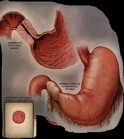
Gastric outlet obstruction is a disorder wherein there is an obstruction in the opening of the stomach (Pylorus), blocking the entrance of ingested food coming from the stomach to the duodenum.

Pyloric stenosis pics

Gastric outlet obstruction showing Pyloric stenosis

​Gastric Outlet Obstruction Symptoms

  • Vomiting – Vomiting is a characteristic clinical symptoms. Usually right after eating, output is either yellow or green. It also contains foods eaten more than 12 hours earlier.
  • Epigastric pain – Usually felt in the upper part of the abdomen, accompanied by acute pain that worsens.
  • Constipation – Passing of stool is difficult and painful due to dry and hardened feces.
  • Loss of weight – Sudden weight loss occurs due to fluid loss and poor appetite.
  • Hypokalemia and metabolic acidosis – Due to vomiting, there is a loss of electrolytes like potassium and may lead to metabolic acidosis.
  • Satiety
  • Feeling of heaviness especially after meal
  • Difficulty and painful swallowing of food
  • Abnormal enlargement of the stomach – Hypertrophy of the muscles in the stomach may be present because of obstruction.

​Gastric Outlet Obstruction Causes

Peptic ulcer disease

PUD is the most common cause of Gastric outlet obstruction, approximately 60% to 65% of total cases. The obstruction often occurs in the duodenum, and sometimes in the pyloric channel, prepyloric gastric antrum, and in rare cases, in the body of the stomach. Peptic ulcer disease is followed by narrowing of the lumen and the outcome is sudden contraction of the stomach muscles, inflammation and edema.

Malignant Tumor

The second most common cause of gastric outlet obstruction, for about 30% to 35% of all cases. Lesions usually appear near the pylorus, symbolizing carcinoma of the antrum and it grows circumferentially.

Benign Tumor

In rare cases, benign polyps in the pylorus can cause gastric outlet obstruction. The polyps are pushed into the duodenum through peristalsis.

Inflammatory diseases

Inflammatory disorders can cause accumulation of excess amount of fluids and sudden muscle contraction that may lead to gastric outlet obstruction. Crohn’s disease commonly develops obstruction. Others are Sarcoidosis, Tubercolosis and Syphilis has been reported to be contributing factor in thickening of the gastric wall that may lead to obstruction. Pancreatitis and cholecystitis can stimulate inflammatory contractions that may result in the destruction of proximal duodenum lumen and gastric outlet obstruction.

Organoaxial volvulus

Twisting of the stomach on itself is also one cause of gastric outlet obstruction.

Ingestion of Foreign Body

This happens very uncommonly. Unintentional among many, but in patients who are mentally impaired, they commit it intentionally.

Diagnosis

Physical Assessment

Physicians initially perform physical examinations on the suspected patients. Auscultation and percussion of the stomach are done by the physician as part of the assessment. There can be a palpable lump in the epigastric region  or a splashing sound can be heard in a dilated stomach.

Barium Meal

Ingestion of barium sulfate followed by x-rays to visualize the stomach and duodenum. It reveals positive abdominal distention and narrowing of the pyloric canal.

Endoscopy

It is a noniinvasive procedure that allows inner visualization of the upper gastrointestinal tract. The esophagus, stomach and duodenum are the involved. This procedure is used to detect gastric tumors and ulcers. Gastric juices can be also collected to be examined for malignant cells.

Abdominal ultrasound

The shadow image on the ultrasound displays distension and enlargement of the stomach.

CT Scan with contrast

Imaging through computed tomography with contrast displays tumors and obstructions in the stomach.

Electrolytes

Monitoring of electrolyte levels such as Potassium, Sodium, and Chloride. Imbalances in these electrolytes may occur due to continuous vomiting and dehydration.

Treatment

1. Proton pump inhibitors

These drugs are used to treat peptic ulcers. They decrease the secretion of gastric acid.

2. Nasogastric suction

A procedure wherein the gastric contents inside the stomach are drained. Using a plastic tube inserted in the nose to the small intestine to remove secretions. However, it is contraindicated among those with esophageal obstruction and blocked airway.

3. Electrolytes replacement

Electrolyte replacement is done to replace what is lost and to maintain the normal level and prevent complications.

4. Antrectomy

Surgical removal of the Antrum, part of the stomach just inside the pylorus.

5. Vagotomy

A surgical procedure that involves cutting one or more branches of the Vagus nerve. The Vagus nerve has a major role in producing and regulating gastric acid secretions. It typically reduces the rate of gastric secretion, usually used in treatment of a peptic ulcer.

6. Billroth I

It is the removal of the Pylorus and the proximal stomach is connected to the duodenum. For the treatment of peptic ulcer and gastric cancer

7. Gastrojejunostomy

Also called Billroth II, it is a surgical procedure that creates a connection between the jejunum (the middle part of the small intestine between the duodenum and ileum) and the stomach. This allow gastric contents to directly enter the jejunum and for proper food absorption.

8. Parenteral Nutrition

Supplementary nutrients via Total Parenteral Nutrition (TPN) or Distal feeding tube are beneficial among patients with gastric outlet obstruction since their nutritional status gets worse.
No responses yet

Seroma – Definition, Symptoms, Fluid, Treatment, Types

Mar 20 2014 Published by under Diseases & Conditions

What is Seroma?

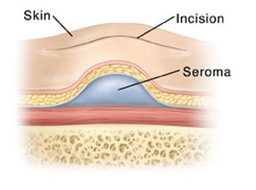
A Seroma is a collection of serous fluid that develops after surgery or any blunt injury. Serous fluid is the fluid compartment of blood that seeps out when blood vessels are injured or ruptured during operation. Inflammation on the surgical site also increases the capillary permeability, thus increasing the flow of plasma from blood vessels into the interstitial spaces (spaces between cells).

Seroma appears like a bump or swelling on the surgical site and may contain clear to yellowish fluid. Surgeons consider seroma as a post-surgical injury that needs proper medical treatment, although seromas are not considered life-threatening.

Seroma Formation

Seroma forms several compartments in the surgical site. These are formation of a seroma cavity with seroma fluid inside.

Seroma Cavity

A thin epithelium usually covers the seroma fluid and resembles a small cavity like a cyst. It usually contains cells around it which may continue to produce serous fluid. In addition, fibrin clots may form on the cavity and encapsulates the accumulation. Some seroma needs surgical excision of the cavity because this cannot be removed by draining.

Seroma Fluid

Inside the seroma cavity is a serous fluid accumulation that originates from blood vessel injury. Over time, fibrin clots form around the fluid and result in a cavity. Fibrin clots develop because the blood contains large amounts of clotting factors and platelets responsible for clot formation.

Seroma Fluid Contents

Seroma fluid contains various substances. These are:

  1. Serum

The basic content of seroma fluid is serum, the fluid component of blood. However, in one study regarding minimally invasive seroma removal, it was found out that other substances are present, including:

  1. Fibrin Clot
  2. Fibrous tissue
  3. Hematoma
  4. Lymphatic fluid

Hematomas and lymphoceles are different from seromas, however, if the injury is severe, hematoma and lymph may be present inside a seroma cavity.

Types

Types of seroma depend on the specific cause. The following bullets outline the most common types of seroma:

Surgery-Related Seroma

These are serous fluid accumulations as a result of injury to blood vessels following a surgery. These are often uncomplicated; however, medical treatment should be done to prevent enlarging of the seroma cavity.

Infected Seroma

Certain injuries may also cause seroma. Dirty injuries (such as an abdominal puncture of non-sterile equipment, motor vehicle accidents, etc.) or those injuries that are infected may also lead to an infected seroma. Aside from draining, this requires antibiotic therapy.

Plastic Surgery-Related Seroma

People who undergo aesthetic surgeries such as tummy tucks or facial plastic surgery may potentially lead to seroma formation.

Seroma at Various Places

Seroma can develop in various places on the body depending on the site of injury or surgery. The most common sites for seroma development are located in the following:

  1. Breast
  2. Neck
  3. Abdomen
  4. Subcutaneous areas
  5. Axilla
  6. Inguinal Area
  7. Lumber Area

Seroma Causes

Various factors contribute to the development of seroma. These include:

Abdominal Surgery

The most common cause of seroma is major abdominal surgery because of the extensive blood supply in the area. Poor use of sutures and antiseptics usually lead to seroma formation.

Mastectomy is the surgical removal of the breast. It is also one of the most common causes of seroma formation. When malignant tumors are removed, the tendency is to injure adjacent blood vessels, leading to inflammation. Inflammatory response causes plasma to leak out and accumulate in a cavity. Removal of the adjacent lymph nodes on the axilla also increase the risk of developing cavities. In addition, removal of the breast tissue leaves an empty space where it was originally. The walls around it may cause leakage of serous fluid and further lead to seroma

Hernia Repair

Hernia repairs cause seroma formation when not properly carried out. Seroma forms on areas near the hernia such as in the inguinal area or abdomen.

Hysterectomy

Seroma after hysterectomy is uncommon, but it may develop as a result of incompetent surgeons handling the operation.

Plastic Surgery

Seroma formation in the neck is common after facial plastic surgeries. A drain is usually placed to prevent the accumulation of fluid after surgery.

Cesarean Section

Just like in other abdominal surgeries, seroma is seen in patients undergoing C-section.

Partial Breast Radiation Therapy

Radiation therapy on the breast has been found recently to cause development of seromas.

Other Injuries

When the body is traumatized or injured, inflammatory response occurs and brings about seroma formation. Just like surgeries, any type of injury is a potential reason for seroma development. Documented injuries that have led to seroma are falls and vehicular accidents.

Seroma Symptoms

Symptoms of seroma are easily accessed and differentiated from other fluid saccumulations. The symptoms include:

Swelling

Accumulation of fluid under the skin causes swelling to be noticed in the area affected

Inflammation

Redness, warmth, and pain is usually seen in the surrounding areas

Palpable Fluid Movement

When the overlying skin is palpated, a fluid wave may be felt as a sign of fluid accumulating under it.

Calcification

When seroma heals, it leaves a calcification or hardening of the skin overlying it.

Infection

In cases of infected seroma, fevers, chills, and pus formation may occur.

Diagnosis

Seroma diagnosis is focused on imaging studies to confirm the presence and location. These include CT-scans and ultrasound. Prior to the diagnostic test, a thorough physical examination is done by the surgeon or physician.

Seroma Treatment

Seroma may be difficult to treat when infection or complications arise. It usually takes time to heal to about several weeks. Treatment of seroma is jointly undertaken by the medical and surgical team. These include:

Drainage

The primary management of seroma is draining of the accumulated fluid. This is done by surgeons where they insert a surgical needle on the site to draw fluid. Some patients may need the drainage several times to completely remove the accumulation. Some large seromas also need a temporary drain with bulb to continuously remove fluids.

After fluid aspiration, physicians subject the fluid to pathologic analysis to check if there is blood, pus, or other components. This may add additional concern and may need additional treatments.

Introduction of needles and puncture of the skin may introduce infection, so sterile techniques are used throughout the draining.

Preparation for the procedure includes NPO (Nothing per Orem) orders, especially when the abdomen is to be drained. Bowel and bladder preparation is also instructed to prevent accidental puncture of these areas due to distention. Patients are informed that the procedure will be uncomfortable but not painful because of local anesthetic administration.

Surgery

In serious cases of seroma, draining is not enough to remove the fluid. In some cases, the seroma cavity needs to be removed because it may still continue to produce fluids despite aspiration. An open surgery is done and the seroma cavity is surgically excised and sent for biopsy to determine any malignancies.

Antibiotic treatment

Infection may develop after a long withstanding seroma. When pathologic studies confirm presence of pus in the fluid, then infection has occurred. In this case, antibiotic treatment is needed to eradicate the infection. The most common antibiotics given are penicillins, cephalosporins and macrolides. It may be administered by mouth but extensive infections may need re-hospitalization and intravenous antibiotic administration.

Newer Treatment Studies

Research studies are trying to confirm if administration of steroids after surgery is essential in the prevention of seroma. However, careful administration should be ensured as these drugs may cause a lot of side-effects including immunosuppression.

Healing Naturally

When seromas are not extensive or too large, sometimes doctors suggest letting it heal naturally without surgery or draining because it may be reabsorbed by the body. Natural healing may vary from person to person and may heal in a few weeks, months, or even in a year. Seromas are not fatal conditions, but complications may be serious. Medical consultation should be done if fever, severe pain, and leaking of fluid in the skin develops.

Complications

Complications of seroma are related to healing of the wound. Because of accumulation of fluids on the surgical site, blood circulation is impaired, thus preventing white blood cells and other reparative substances to go to the site of injury. In cases of a very large seroma, it may impair the approximation of sutures, leading to dehiscence and evisceration (opening of the wound). In the long run, delayed wound healing attracts the proliferation of bacteria and leads to infection.

Seroma vs. Hematoma

Seroma is sometimes confused with hematoma and others such as lymphocele and abscesses. Seroma is the accumulation of serous fluids (usually clear), while hematoma formation is a localized accumulation of red blood cells (from the word “heme” which means blood). Moreover, lymphocele is the accumulation of lymph, and abscess is the accumulation of pus. All of these are characterized by swelling and inflammation so definite diagnosis is needed to arrive at an appropriate treatment method.

Seroma Pictures

anatomy of seroma

Image 1 – Anatomy of seroma

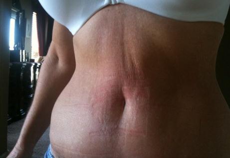
Seroma After plastic surgery

Image 2 – Seroma after plastic surgery (after Tummy tuck)


No responses yet

Sweaty Feet – Causes, Remedy, How to Stop & Levels

Mar 12 2014 Published by under Diseases & Conditions

What is Sweaty feet?

Hyperhidrosis is a medical term for excessive sweating that is not normal or usual. It can be generalized or localized. General Hyperhidrosis affects the entire body and is usually caused by an underlying condition, commonly by infections or by endocrine or neurological disorder.

Sweaty Feet Image

Localized Hyperhidrosis involves a particular part of the body. It includes the feet (Plantar Hyperhidrosis), hands (Palmar hyperhidrosis), armpits (Axillary Hyperhidrosis) and groin because of active sweat glands in the following areas. The most frequently affected area is the armpits, hands and feet.

Excessive sweating of the feet or having sweaty feet can be embarrassing and disturbing in a person’s daily activities. Excessive perspiration can attract bacteria that results in foul-smelling odor of the feet and other conditions like Athlete’s foot. Therefore, early detection and management is important to prevent further complications.

Sweaty feet affects both men and women, most commonly between the ages of 25 to 64, but it can also start in early childhood.

Sweaty Feet Levels:

Mild Sweating

The soles are very moist and perspire much more than others, has mild undesirable odor and discomfort.

Moderate

Drops of sweat are present, but sweating is confined to the feet and occurs frequently. Mostly, it occurs 4 to 6 hours after taking a bath or shower. The skin is prone to blisters and swelling.

Severe

The sweating is immoderate, socks and shoes worn are wet. The sweat drips off the feet. The undesirable odor is intense. Blisters and peeling of skin are more common.

Sweaty Feet Causes

1. Idiopathic

The main reason of excessive sweating of the feet in still unknown.

2. Hyperfunctioning of Autonomic Nervous System

Overstimulation of the neuronal transmission to the sweat glands results in over-secretion of sweat.

3. Unsuitable footwear

Wearing unsuitable socks and shoes like shoes made with plastics do not allow evaporation nor absorption of the sweat, resulting in wet feet.

4. Withdrawal Syndrome

Alcohol and drug abuse is also related in hyper-secretion of sweat. Alcohol and drugs have an effect on the blood vessels, making them dilated and  they raise the body’s core temperature, leading to increased sweating

5. Medications

Drugs like antidepressants, beta blockers and parasympathicomimetics may induce sweating.

6. Hormonal Disturbance

When certain hormones lose their function, it may involve excessive perspiration.

7. Overweight and Obesity

8. Disoder of the Thyroid Glands

  • Hyperthroidism is the most common disease that manifests excessive sweating due to hyperfunctioning of thyroid glands and hypersecrition of hormones.
  • Pheochromocytoma, or Adrenal Cortex tumor – this condition results in hyper-secretion of Epinephrine and Norepinephrine. These two hormones elevate the blood pressure and also stimulate excessive sweating.

 9. Disorder of the Nervous System

  • Nerve injury or damage can cause very unusual perspiration.
  • Encephalitis or inflammation of the brain may also elevate sweat secretions

How to stop sweaty feet?

  • Wear shoes without synthetic lining, or choose those with lined with leather.
  • Allow your shoes to be ventilated after every use. This allows drying before shoes are worn again.
  • Choose socks of man-made fibres because they can absorb and hold sweat very effectively. Socks must be exactly fitted, not too tight, to allow proper ventilation and air transport.
  • You can try foot antiperspirant like Aluminum chloride solution. Apply it at night, allow it to dry and rinse off in the morning.
  • Consult a doctor when infections occur, this is to treat immediately and prevent further complications.

Sweaty Feet Treatments

1. Iontopheresis

This is a treatment done by physiotherapist in hospitals. The feet are placed in a tub of water in which there is a very minimal amount of electrical currents passing. This will last for 30 to 40 minutes. It is non-invasive and does not involve surgical procedure.

2. Anticholinergics

These medication works with the nerves in-charge in sweating to reduce and prevent excessive secretion of sweat.

3. Antiperspirants

One common antiperspirants is Aluminum Chloride which decreases the secretion of sweat by obstructing the pores of the eccrine glands.

4. Botulinum Toxin injection

Botox injections block the nerves within the palms and soles. It is an invasive procedure that can be performed with just little amount of anesthesia.

No responses yet

Ganglion Cyst – Pictures, Wrist, Foot, Treatment, Surgery Removal

Mar 05 2014 Published by under Diseases & Conditions

What is a Ganglion Cyst?

We are living in a generation where cancer is one of the leading causes of morbidity and mortality; therefore, it is normal for us to panic whenever we notice any lump or tumor in our body. However, several lumps that occur are usually benign, like Ganglion cyst. Ganglion cysts, also known as Bible cysts, are a soft tissue mass in the joints.

How Ganglion Cyst formed?

A ganglion cyst can be described as a tumor or swelling on top of the joint or on the tendon itself. It appears like a sac of liquid which can be firm or spongy depending on the size. The liquid that fills the cyst is a sticky, thick, colorless, and jelly-like material. When the synovium in the surrounding tendon degenerates, the tendon sheath tends to buckle and weaken which causes the synovium to swell and form lumps.

Signs & Symptoms

The signs and symptoms of ganglion cyst include:

  1. Freely movable mass over a joint
  2. About 1-3 cm in size but may grow bigger
  3. Localized pain exacerbated by dorsiflexion of the wrist or moving the joint, but some cases are painless
  4. Swelling around the area which indicates compression of circulation
  5. Weakness or numbness on the distal portions such as the fingers which may indicate nerve compression

Characteristics

  1. Ganglion cyst is a type of completely harmless tumor.
  2. Ganglion cysts are more common in the female population.
  3. In addition, people between 20 – 60 years of age are more at risk for ganglion cysts, which accounts for seventy percent of cases.
  4. An individual can have one large cyst or multiple smaller cysts that arise from one cyst which project a grape-like appearance.
  5. Unlike cancerous tumors, ganglion cysts are mostly made up of soft tissues in the body and not toxins.
  6. Ganglion cysts are not malignant and do not spread to other areas of the body.
  7. Ganglion cyst may be painful and painless.
  8. Small lumps are usually painless, however as the size increases, it may compress some of the nerves and blood vessels and cause pain.

At Wrist, Foot & Ankle

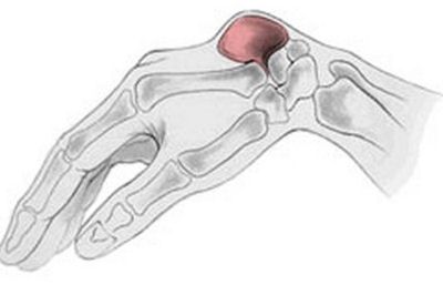
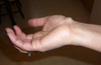
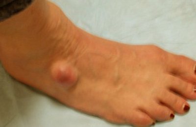
Ganglion cysts are most likely to develop on the wrist joints, the back of the hand, and the anterior portion of the wrist. They can also develop on the knees, ankle, and top of the foot.

Ganglion cysts on the wrist, foot, and ankle arise from the accumulation of non-cancerous fluid which results to a palpable lump or tumors. These fluid-filled capsules have a tendency to increase in size, but they don’t have the ability to multiply. Although there are some cases of multiple cysts, it usually originates from a single main tumor.

Causes

The most common risk factor for ganglion cyst development is trauma.

Trauma

Ganglion cysts are commonly caused by trauma to the wrist tendon. After trauma, there could be a gradual or sudden onset of symptoms. Injury may involve blunt trauma to the area or repetitive movements such as frequent dorsiflexion and plantar flexion of the wrists as seen in typists. Injury to the joint causes the synovial cavity to break which eventually leads to lump formation. Injury to the tendon covering may also cause tissues to bulge and form lumps.

Diagnosis

Every palpable lump or tumor in the body needs careful inspection and examination. Physical examination is done to assess the swelling and tenderness around the area. Definitive diagnosis involves excision of the cyst and submitting it for histological examination or biopsy to determine malignancy if any. Almost all of cases of ganglion cyst are benign.

Treatment Options

Ganglion cysts are managed primarily through removal of the lump. The following are possible methods of removing the ganglion cyst or relieving symptoms:

Surgery

Surgical removal of the ganglion cyst is the most common procedure to completely remove the cyst and prevent recurrence. The procedure involves minor operation to excise the cyst or cysts in the wrist. The excision also involves the removal of the cyst capsule to avoid new cysts from growing.

The procedure starts from injecting local anesthesia over the area to prevent pain sensation. Then, a small incision is made with the insertion of an arthroscope to visualize the inside portion of the joint. Upon confirming the location of the cyst, a small instrument is inserted to remove the lump.

The procedure is done in an out-patient setting and the surgical incision usually heals in a matter of one to two weeks.

Aspiration

Aspiration is a more conservative approach to ganglion cyst removal. The procedure involves splinting the wrist, followed by application of a local anesthetic. A small needle is inserted then the fluid is aspirated. Aspiration should be slow because sudden pulling of the plunger may cause further trauma inside the joint.

After aspiration, steroids are injected to prevent inflammation. Aspiration is effective in removing the cyst but recurrences are likely.

Non-steroidal Anti-inflammatory Drugs

NSAIDs such as ibuprofen, naproxen and aspirin may be taken to reduce inflammation and pain.

Natural Remedies

Remedies may be instituted to decrease pain and inflammation. Warm compresses may be placed over the area for 15 minutes three times a day to relieve the condition.

Myths regarding management of ganglion cyst should be ignored because these may lead to further injury. Some people suggest the use of a Bible to smash the lump to be removed. Of course, this is a primitive practice which made ganglion cysts known as Bible cysts. Also, avoid self-pricking the lump to prevent infection.

Complications

Although ganglion cysts are benign, it may lead to possible complications which are usually not serious. Complications are often prevented through removal of the cysts that may be bothersome. Complications include:

Temporary loss of wrist function

Because of the swelling and nerve compression, the wrist may not be able to perform its functions, especially when the cysts grow big. Numbness and pain also result in poor movement of the hands and wrists.

Severe Pain

Significant pain may be experienced because of continued pressure on the nerves which stimulates nociception or pain sensation.

Infection

Infection may arise as a complication of ganglion excision. This may be prevented by ensuring aseptic technique throughout the surgery.

Prognosis

Since ganglion cysts are benign, there is a good prognosis. Surgical excision or aspiration of fluid can easily remove the cyst. Although chances of recurrence are high, patients should not worry because the recurrences are also benign. Recurrences are likely if the capsule that surrounds the cyst is not removed.

Prevention

Ganglion cysts may be prevented by avoiding excessive stress on the wrist joint. People should engage in an active range of motion exercises in a day to allow the circulation over the area. People who are likely to move the wrist most of the time for work, such as typists, construction workers and others, should ensure to rest the wrists after heavy work.

Ganglion Cysts Pictures

Pictures, photos, images of ganglion cyst at wrist, ankle and more.

bible cyst on wrist (diagramatic)

ganglion cyst at wrist region

ganglion cyst at wrist of upper arm

ganglion cyst at front of wrist region

ganglion cyst at foot

ganglion cyst at foot (ankle)

No responses yet

Furuncle – Definition, Treatment, Pictures, Causes, Symptoms

Mar 01 2014 Published by under Diseases & Conditions

What is Furuncle?

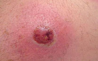
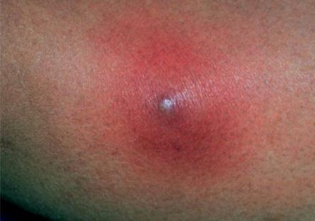
Furuncle is a skin infection surrounding the follicles of the hair, also termed as boils. It is a bacterial infection that results in swelling of the skin around the hair follicle with resulting pus and tissue debris formation. This condition is known as an abscess formation where pus accumulates beneath the dermis and extends to the subcutaneous area. When multiple hair follicles inflame and fuse, they are known as carbuncles. Reccurring boils is termed as furunculosis

Furuncle Causes

Main Cause

The main source of furuncle is a bacterium called Staphylococcus aureus. It is a gram positive, round microorganism which is present in most of the areas of the body, specifically the skin. It is a transient micro flora in the skin which does not cause significant illness when there is no open wound. In furuncles, when the hair follicles are exposed such as in removal of a hair shaft, microorganisms tend to enter and proliferate, causing infection. Since they are an opportunistic bacteria, they also multiply and grow when the immune system weakens.

Other causes and risk factors

The Tumbu fly in Africa causes myiasis which results in furuncles. Aside from this, underlying conditions such as diabetes mellitus, malnutrition, anemia, neoplasms, intake of immunosupressants, and antibiotic overuse increases the risk for developing furuncles because of growth of opportunistic microorganisms such as S. aureus.

Furuncle Signs and Symptoms

Manifestations of boils include both local as well as systemic signs and symptoms.

Local Manifestations

Local signs and symptoms of furuncle include:

  1. Pustules (contains pus), pea-sized lesion located on the hair follicle. Advanced infection may be as large as a ping-pong ball
  2. Redness, swelling, and warm skin around the infected site
  3. Whitish to yellowish pinpoint on top of the boil can be observed when it is ready to drain pus.
  4. Mild to moderate pain on the lesion

Systemic Manifestations

When furuncles are severe, they tend to produce systemic sign and symptoms as a sign of spread of the infection. These include:

  1. Fever
  2. Lymph node swelling, especially the ones adjacent or near the furuncle
  3. Body malaise and fatigue
  4. Itching

When furuncles drain, the lesion opens and a whitish to yellowish pus oozes out. Once the infection is resolved, the lesion dries and turns into scab and leaves a dark spot when completely healed within two weeks.

Furuncle Diagnosis

Diagnosis is done through culture and sensitivity tests. A culture is often done by getting a sample of the drain to allow the bacteria to grow and replicate. Once it has been achieved, sensitivity tests to several antibiotics are done to determine the most appropriate medication to be given where the bacteria is most sensitive.

Furuncle Treatment

Although furuncles may heal on their own as long as further infection is prevented, some cases need to be managed by health practitioners. It is best to consult physicians to select appropriate treatments and recommendations. Treatments of furuncles include medical and surgical as well as a supportive approach.

Medical Management

Oral Antibiotic therapy

Antibiotics are the primary treatment for furuncles since it is an infectious disease. The most common antibiotics used is penicillin. These are the first line of drugs for furuncles, however, patients should take the medication as prescribed (seven to ten days depending on the severity of the infection) to prevent drug resistance. Methicillin Resistant Staphylococcus aureus (MRSA) is a severe, life-threatening infection as a result of non-compliance to antibiotic therapy. Antibiotics also need to be administered by mouth or intravenously. Local application such as sprinkling of antibiotic powders is not effective because absorption is not optimum.

Topical application of antibiotics.

Some antibiotics are prepared as creams and ointments. These are an approved and prescribed way of applying local antibiotics rather than sprinkling powder from antibiotic capsules. The boil needs to be washed thoroughly with soap and water before topical application of creams.

Surgical Management

Perform Incision and Drainage

Surgeons usually perform incision and drainage to open the lesion and lremove pus and other debris. This should be done by medical professionals only because doing this at home can cause more infection. Sterile equipment is used in the hospital or clinics to prevent further infection. After the surgical procedure, antiseptics such as iodine or antibiotic creams are applied to kill transient microorganisms. A bandage may be placed to prevent further entry of bacteria and other microorganisms in the wound and promote healing. Bandages should be replaced every day aseptically. Physicians and nurses usually provide home instructions on how to replace the bandage.

Supportive management

Furuncles should be continuously managed at home. In some cases where patients are unable to consult health practitioners, home management can be instituted to reduce symptoms. However, consultation should be done whenever possible. Home management includes:

Apply warm packs over the affected area

A warm compress applied over the furuncle helps to drain the pus and hastens healing. Do this by using a soft cloth soaked in warm water. Do not open the wound by other means and never squeeze out the pus. Physicians should be the one to do that to prevent reentry of bacteria. Warm packs also sooth the skin and reduce pain.

Clean the infected area

Always clean the boils with soap and water and other antiseptic washes to reduce bacteria. Also, apply antiseptic creams as prescribed. Never place erosive and irritating chemicals such as alcohol to prevent further injury.

Apply dressings.

Use dressings to cover the boil and change them every day. Properly dispose of the infected article to prevent spread of infection.

Hand washing

Wash your hands before and after contact with the lesion to prevent spread of bacteria.

Never re-use towels and never borrow and lend them to others.

Re-using towels will just bring the bacteria in contact with the skin again and spread it to other areas of the body. Borrowing and lending personal articles such as clothes and towels can lead to cross infection.

Boils usually heal on their own when they are not complicated. Healthcare practitioners should be consulted when the boil is too deep or too large, boils last more than two weeks, systemic manifestations are present, for recurring boils, and any boil present on the face and back.

Furuncle Complications

Staphylococcus aureus may spread to other areas of the body, causing more serious complications such as:

Spinal cord, kidney, and brain abscess

Infection may reach vital organs and cause infection in the area.

Osteomyelitis

Bones are one of the potential areas of spread due to blood circulation. When osteomyelitis occurs, the patient may have severe bone diseases and fractures.

Pneumonia

The lungs are easily infected because Staphylococcus aureus are always present in the nostrils. Any furuncle in the area may descend to the lungs.

Endocarditis

When the heart muscle (endocardium) becomes infected, myocardial contractility may be impaired and lead to circulatory problems.

Sepsis

Bacteria may spread and multiply in the blood stream, causing sepsis.

Increased severity of other diseases

Presence of S. aureus may intensify effects of toxic shock syndrome, food poisoning, septic shock, and burns.

Furuncle Prevention

Since furuncles are primarily due to infection, it is an easily-preventable disease. The following prevention strategies are essential in avoiding furuncles and other infections.

Proper hygiene

Thorough hand washing and bathing every day helps in reducing S. aureus. It also prevents clogging of hair follicles and the spread of infection.

Antimicrobial soaps

Use soaps with antimicrobial properties to kill potentially harmful bacteria.

Antiseptic washes and disinfectants

Use alcohol and hand sanitizers when washing is not possible such as in areas with no accessible water and soap. If wound is present, clean it with antiseptics such as povidone-iodine to prevent infection.

Furuncle Pictures

Furuncle photo

furuncle

Furuncle photo

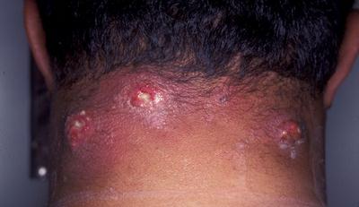
Image 3 – Furuncle At nape of neck

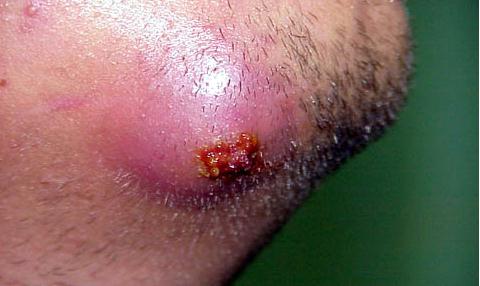
furuncle at chin

Image 4- Furuncle at chin

No responses yet

© 2017 HealthFoxx.com. All Rights Reserved. Privacy Policy
This website is for informational purposes only and Is not a substitute for medical advice, diagnosis or treatment.