Archive for July, 2013

Maple Syrup Urine Disease – Pictures, Symptoms, Treatment, Diagnosis

Jul 25 2013 Published by under Diseases & Conditions

What is Maple Syrup Urine Disease (MSUD)?

Maple Syrup Urine disease is a congenital condition characterized by a metabolic error involving branched-chain amino acids. The metabolic error results in urine with a distinctive sweet smell.  It is also an autosomal recessive disease, which means the condition can be inherited.

What Happens When an Infant has Maple Syrup Urine Disease (MSUD)?

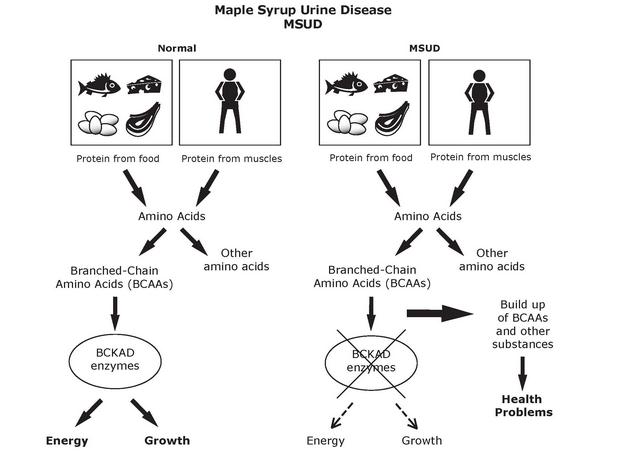
MSUD is a condition characterized by a faulty metabolism of branched-chain amino acids. Amino acids are the building blocks of proteins; proteins in turn are the building blocks of almost everything in the human body. Proteins are important components of muscles and bones, skin and internal organs, connective tissues, blood components and much more. Without amino acids, protein build up wouldn’t be possible, and without protein, building essential components of life also wouldn’t be possible.

There are three examples of branched chain amino acids: isoleucine, leucine, and valine. These amino acids are considered essential amino acids. They are called “essential” because they are not produced by the body (therefore we must get them from outside sources, e.g. diet) and are important in the regulation of certain body processes.

What happens with Maple Syrup Urine Disease is the body has defective enzymes to break down or catabolyze the branched chain amino acids. Breakdown or catabolism of these amino acids is an essential process so that the body can use the amino acids and also prevent them from accumulating in the body. Because the enzyme responsible for the breakdown is defective (as it cannot breakdown or process the amino acids properly), the amino acids accumulate. The accumulation of the amino acids poses a danger to the brain and overall neural function of the infant as these proteins and their byproducts are toxic. The condition can then lead to progressive degeneration of brain functions, seizures, coma and death, especially when left untreated.

What Causes Maple Syrup Urine Disease

Alterations in genes or gene mutations are said to be the primary cause of Maple Syrup Urine Disease. Changes in the genes DBT, DLT, BCKDHA and BCKDHB cause the condition. These genes are responsible for instructing the cells to effectively process and break down the amino acids isoleucine, leucine, and valine. Because the genes are defective, proper instructions are not given to the cells, leading to a faulty processing of the proteins.

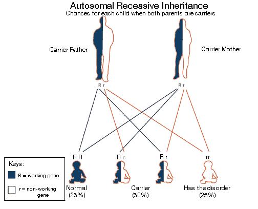
The gene mutations are inherited conditions and the disease follows an autosomal recessive pattern. This means each cell has both copies of the faulty gene. Parents of a person with an autosomal recessive disease carry a copy of the faulty gene, but most often the parents don’t manifest the signs and symptoms of the disease.

Symptoms of Maple Syrup Urine Disease

Maple Syrup Urine Disease (MUSD) symptoms depend on the extent or the type of MUSD they have. The signs and symptoms of MUSD according to its classification are listed below.

Classic Maple Syrup Urine Disease

Classic Maple Syrup Urine Disease is the most common and most severe type. The condition is present immediately or after 4 to 7 days from birth; symptoms of Classic MUSD are as follows:

  • Vomiting
  • Poor feeding
  • Delay in breast feeding
  • Problems in weight gain
  • Progressive lethargy
  • Muscular hypotonia and hypertonia (alternating)
  • Dystonia
  • Encephalopathy
  • Seizures
  • Pancreatitis

Intermediate Maple Syrup Urine Disease

This is a rare type of MUSD where symptoms can present themselves at any age. Signs and symptoms of this type of MUSD include:

  • Delay in the development
  • Neurological impairment
  • Presence of seizures

Intermittent Maple Syrup Urine Disease

This ranks as the second most common type of MUSD. In this type children develop normally both physically and mentally, however signs and symptoms of MUSD appear when the patients experience catabolic stress and intercurrent conditions such as otitis media. Signs and symptoms of this type include:

  • Lethargy
  • Ataxia
  • Coma
  • Seizures

Thiamine Responsive Maple Syrup Urine Disease

Is another rare type of MUSD where patients respond to thiamine. Neurological signs and symptoms such as lethargy, seizures, ataxia, are present.

E3-deficient MSUD

This type is a very rare form of Maple Syrup Urine Disease. The signs and symptoms are almost similar to “intermediate” except that this type is accompanied by lactic acidosis. Other deficiencies of the disease include:

  • Pyruvate
  • BCKD
  • Alpha ketoglutarate dehydrogenase complex

How Is Maple Syrup Urine Disease Diagnosed?

Maple Syrup Urine Disease can be diagnosed through the following:

1. Examination of clinical features

Physicians examine the infant or the child for neurological problems, and other signs and symptoms reflective of MUSD. The classic sign of MUSD is the sweet scent of urine.

2. Blood Enzyme Tests

Enzymes present in the blood are also tested to diagnose MUSD. Decreased levels of the enzyme BCKAD and an increase in BCAA are usually suggestive of the condition.

3. Molecular Gene Testing

This diagnostic test aims to identify the three genes associated with Maple Syrup Urine Disease; these genes are BCKDHA, BCKDHB and DBT.

How Is Maple Syrup Urine (MUSD) Treated?

Treatment for MUSD usually involves:

1. Dietary Restrictions

Once diagnosed, strict and permanent dietary restrictions are implemented. Patients must avoid food sources high in leucine and other branched chain amino acids. This can be done by calculating the individual’s tolerance to leucine and choosing food sources that are appropriate to the patient’s tolerance. This is done for the first 6-12 months of life. Strict monitoring and implementation of the diet is the mainstay treatment for MUSD.

2. Glucose Infusions and Insulin

Glucose infusions are done to manage metabolic decomposition. Insulin is also given to manage or promote anabolism.

3. Liver Transplants

This treatment is done on patients with the classic form of MUSD. A liver transplant may halt the progressive decline in neurological function patients have, but the procedure does not reverse the existing damage patients have suffered.

Pictures

Maple Syrup Urine Disease image

Maple Syrup Urine Disease photo

No responses yet

Fibrodysplasia Ossificans Progressiva – Pictures, Symptoms, Treatment

Jul 22 2013 Published by under Diseases & Conditions

What is Fibrodysplasia Ossificans Progressiva?

Fibrodysplasia Ossificans Progressiva is a genetic disorder that affects the muscular and connective tissues in the human body.  It is responsible for body movements that then transform the connective tissue into bone, called ossification. When ossification occurs, malformation of a bone in the external area of the skeleton will take place.  Also called the extra-skeletal or heterotopic bone, it is confined to movement in the spine, chest, neck, shoulders, wrists, elbows hips, knees, elbows hips, ankles and jaw but does not restrict progression.

FOP is an atypical disorder because it only affects one in two million people; males and females are both affected in an equal manner. Surgery is prohibited and it can be dangerous to even fall down, because this may cause swelling and inflammation in the muscle and may intensify rapid ossification in the area that is injured.

Fibrodysplasia Ossificans Progressiva  Pictures

Symptoms
Symptoms of Fibrodysplasia Ossificans progressive may include:

  1. Vagus Deviation, a malformation in an outward appearance of the great big toe, the hallmark symptom found in newborns.
  2. During the first 10-20 years of life, continuous loss of mobility as the joints become affected and growths develop from the jaw, neck down to the spine, thoracic area, knees, and feet, just as bones would grow in a fetus.
  3. Difficulty or inability to fully open the mouth, causing a difficulty in speaking and eating. This may lead to malnutrition as the person afflicted cannot chew without causing a growth.
  4. Painful flare-ups that last up to 6 -8 weeks. The first flare up that leads to the development of the disorder usually occurs before the age of 10.
  5. Swelling of the tissue and joint stiffness that leads to extreme discomfort.
  6. Some may suffer from low-grade fever.
  7. Development of tumors in the connective tissue and muscle tissue. These lesions lead to formation of bone outside the skeleton called heterotopic ossification in the axial muscles, the fascia, in the ligaments, the tendons, and the joint capsules.
  8. Breathing difficulties as a result of extra bone formation around the rib cage, limiting expansion of the lungs, which leads to TIS (thoracic insufficiency syndrome)

Causes

Germline mutation – Fibrodysplasia Ossificans Progressiva is a genetic disorder in which there is a mutation in a gene. The gene mutation is responsible for FOP and it is already present at birth. In the tissues and cartilaginous area of the skeletal muscles the gene is present because it also helps in regulating the development of bones and muscles in the body. It also contributes to the maintenance in cartilage replacement into a bone, which takes place as a person matures. However, mutation of this gene tells the body to continue to grow bones after the fetus is fully developed in the womb, rather than stop once all bones and tissues are fully formed.

Random occurrences – Some cases FOP are not inherited. The mutation starts at the development of an egg, sperm and embryo.

Inheritance – Fibrodysplasia Ossificans Progressiva is inherited through a autosomal dominant allele which means it is passed on by only one parent.  This is enough for the child to develop the disorder. A parent with FOP has a 50% chance that his/her child will have Fibrodysplasia Ossificans Progressiva. However, this disorder is rarely inherited because most people who have the disease are unable to bear a child.

Diagnosis

FOB is a very uncommon disorder and it is oftentimes:

  1. Diagnosed by its signs and symptoms (tumor-like swellings on the neck, head, shoulders, back and Vagus deviation)
  2. 87% are misdiagnosed with other diseases such as Cancer, aggressive juvenile fibromatosis, and progressive osseous heteroplasia.

Treatment

  1. Early detection is the best way to lessen the progression of the disorder, but not to treat the disease. Surgical removal of the excess bone is not advisable because it may result to more bone formation. There are no effective medical therapy known for FOP; bisphosphonates and corticosteroids are beneficial only during the episodes of flares.
  2. Lessening the progression by avoiding bumps and falls.
  3. There are different medical measures to increase pulmonary function, decrease respiratory compromise, and prevent pneumonia and influenza.  These are all helpful in decreasing the number of deaths from thoracic insufficiency syndrome.
  4. Rehabilitation approaches should be focused on improving daily activities. Occupational therapy and vocational education consultations may be extremely useful for FOP patients.

Scientists discovered in 1999 that squalamine in sharks that might be useful in treating those individuals who are suffering from FOP. Squalamine is an antiangiogenic and can prevent the growth of blood vessels in cartilaginous tissue, thus preventing bone calcification in sharks. In 2002 they started a trial of squalamine. It was terminated because squalamine is a different compound found in sharks that has no such properties of preventing ossification in humans.

Prognosis & Life Expectancy

Unfortunately, due to lack of treatment, the fact that the disease is incurable and fatal, only management of the symptoms, pain, and discomfort are done using prophylactics.

Fibrodysplasia Ossificans Progressiva is a incurable disease at this time so the life expectancy of individuals who suffer from FOP is poor. According to research, the life expectancy of persons with Fibrodysplasia Ossificans Progressiva is up to 41 years of age, but some have survived until the age of 60. Many FOP patients suffer from thoracic insufficiency syndrome (TIS) so some die before the age of 40-41 years old.

FOP is a progressive disorder it gets worse as a person ages. It differs from person to person in severity and is very unpredictable.

Pictures

Fibrodysplasia Ossificans Progressiva    Fibrodysplasia Ossificans Progressiva images   Fibrodysplasia Ossificans Progressiva pictures  Fibrodysplasia Ossificans Progressiva pics

No responses yet

Hormone Replacement Therapy – Benefits, Side Effects for Men & Women

Jul 14 2013 Published by under Obs & Gyn

What is Hormone Replacement Therapy?

Hormone Replacement Therapy

Hormone Replacement Therapy is a classification of medication given to women and men who experience a sudden drop of either progesterone, testosterone and estrogen hormones, usually experienced during menopausal or andropausal stages. Menopause or andropause is a stage in a person’s life where he or she experiences infertility. Hence, to correct it, hormone replacement therapy puts the hormonal level back within normal range. In addition to that, hormone replacement therapy can either be a combination of progesterone and estrogen or singly estrogen or progesterone. The intake of hormone replacement therapy drugs can prevent health problems and discomfort associated with menopausal and andropausal stages.

Hormone Replacement Therapy Indications

The hormone replacement therapy can be used by either men or women:

For men

Normally, the hormone associated with men is testosterone. Men generally have a high amount of testosterone in their body. This is one of the reasons why men are men in the first place. Testosterone is needed to maintain bulk in the muscular area, bone density, reproductive and sexual function, as well as adequate levels of red blood cells. Testosterone has also been shown to sharpens one’s memory and pump up libido. However, when a man reaches 80 years and above, he will experience a sudden decline of the testosterone, indicating that he is about to enter or is in the menopausal stage of development, which is called andropause for men. Hence, hormone replacement therapy for men is packed with testosterone to aid them in maintaining a normal level of the testosterone in their body and prevent andropause’s side effects.

For women

Normally, women suffer earlier menopause than men. When menopause happens, which occurs in everybody without exemption, there will be an imbalance in the hormones, particularly those dealing with estrogen and progesterone. In addition to that, women usually face a different situation during menopause compared to men. They are susceptible to illness during menopausal stage. Hence, there is a great need to correct imbalance hormones with hormone replacement therapy.

Hormone Replacement Therapy image

Hormone Replacement Therapy Contraindications

Despite the fact that hormone replacement therapy may prove to be beneficial for people who are undergoing either andropause or menopause, there are still contraindications to consider before taking in kind of medical treatment therapy.

Men

The contraindications associated in hormone replacement therapy for men are as follows:

  • Men with prostate cancer should not take hormone replacement therapy, for it may lead to enlargement of the prostate
  • Men with coronary disease
  • Men with high level of cholesterol
  • Men suffering from heart disease or heart failure
  • Men diagnosed with liver disease
  • Men who are complaining of chest pain
  • Diabetic men
  • Men who are allergic to the component of testosterone injections known to have benzyl alcohol

These men will not benefit from hormone replacement therapy treatment management. Hence, they must discuss other alternative ways for treatment from their hormonal imbalance.

Women

The following are contraindications in the usage of hormone replacement therapy in women:

  • Breast cancer in the family history
  • Pregnancy
  • Venous thrombosis
  • Liver disease in the severe form
  • Undiagnosed cause of vaginal bleeding
  • Endometrial cancer
  • Coronary artery disease
  • Ovarian cancer in the family history
  • Gallbladder disease
  • Ductal hyperplasia of the breast
  • Uterine fibroids in the medical history

Women who are either experiencing any of medical conditions mentioned above should consult a physician prior to taking hormonal replacement therapy.

Hormone Replacement Therapy Side Effects & Risks

Hormone replacement therapy is not a one hundred percent perfect solution. It also has side effects and risks that you should consider and keep in mind.

Men

  • Sleep apnea
  • Enlargement of the prostate gland
  • Retention of fluids
  • Acne
  • Shrinking of the testicles
  • Excessive red cell growth
  • Possibility of sperm count reduction
  • Blood clotting
  • Strokes
  • Heart attacks
  • Prostate cancer
  • Benign prostate hypertrophy
  • Depression
  • Aggression
  • Breast enlargement

Women

  • Nausea
  • Mood swings
  • Fluid retention
  • Bloating
  • Stroke
  • Heart attack
  • Gallbladder disease
  • Dementia
  • Headaches
  • Weight gain
  • Darkening of the skin
  • Uterine fibroids
  • Worsening of already present endometriosis
  • Pain in the breast region
  • Breast density increases
  • Endometrial cancer
  • Ovarian cancer
  • Blood clots
  • Osteoporosis
  • Breast cancer

Hormone Replacement Therapy Benefits

Men

Men who reportedly take hormone replacement therapy will report the following benefits from the medication treatment:

  • Memory improvement
  • Moods consistency
  • Increase in energy
  • Increase in their muscular strength, size and weight
  • Libido enhancement
  • Lean body mass increases
  • Reduction of body fat

Women

Generally speaking, the hormone replacement therapy for women has low testosterone and high estrogen and progesterone levels. The following are the benefits:

  • Prevents osteoporosis
  • Lowers the risk for colorectal cancer
  • Decrease risk for heart disease
  • Decrease depression
  • Sleep quality is enhanced
  • Slows the aging process
  • Sexuality and libido is enhanced
  • Preventative of tooth disease
  • Weight loss
  • Able to manage or alleviate the symptoms associated with menopause
  • Quality of life is improved
  • Mood elevation
  • Muscular tone improvement
  • Firmer breasts

No responses yet

Whipple Procedure – Complications, Prognosis, Survival Rate, Pictures

Jul 10 2013 Published by under Gut & Liver

What is Whipple Procedure?

Named after an American surgeon, Allen O. Whipple is a surgery called Whipple Procedure also called Pancreaticoduodenectomy. A Whipple procedure is a surgical operation done primarily for Pancreatic Cancer patients. The procedure is usually done so as to remove an enlargement on the duodenum. The duodenum is the first section of the small intestine, or in layman’s terms, the small bowel. It can be the removal of bile duct or head of the pancreas. It can also be done for patients with chronic pancreatitis or for those patients who have bile duct or pancreatic duct strictures.

Whipple Procedure Picture (diagram)

The physician will explain and illustrate a drawing of the sections of the body involved for the patient to see.

The whole procedure is usually completed in four to six hours.  The patient may possibly need a blood transfusion at some stage in the procedure. The surgery can include detaching one or more of the following:

  • Gallbladder
  • Section of the bile duct
  • The duodenum
  • Head of the pancreas
  • Inferior section of the stomach.

Whipple Procedure for Pancreatic Cancer

  • The Whipple procedure is a possible choice for patients who have pancreatic cancer but only if the cancer has not spread past the pancreas. This is an extensive and difficult surgery that is intended to eradicate the entire malignant cells or cancer cells.
  • It includes detaching the head of the pancreas, normally located deep within the abdomen.
  • Additionally, a segment of the bile duct, the gallbladder and the duodenum are also detached.
  • Afterward, the surgeon needs to put back the pancreas, the intestine and the remaining bile duct, called “the reconstruction”.
  • The reconstruction will permit the patient to consume food and digest food just as before the surgery.
  • Usually, a Whipple procedure it will include the surgical removal of a section of the stomach. If the stomach during the Whipple procedure is not removed, then the surgery is called the Pylorus-Preserving Whipple.

Complications

Pancreatic Fistula

Pancreatic fistula can be a complication of Whipple procedure. This happens if there are problems during the healing process of the suture that connects what remains of the pancreas to the intestine.

Leakage of pancreatic juice

Subsequently, leakage of the pancreatic juice can occur. In most cases, the leakage will repair on its own without requiring another surgery. However, in rare cases, another operation might be needed to repair the leak.

Stomach paralysis

Furthermore, 25 out of 100 patients can have stomach paralysis after the procedure. However, one normally recovers in four to six weeks time. In cases of stomach paralysis, the patient is required to temporarily obtain nutrition through a feeding tube.

Side Effects

The usual long term side effects of a Whipple procedure are problems with digestion, such as:

  • Diabetes
  • Malabsorption
  • Weight loss.

Diabetes
After the surgery, diabetes may manifest in the patient who has undergone the surgery. Diabetes is a metabolic problem wherein the body does not produce insulin or lacks insulin to control blood glucose. This can happen in Whipple procedure patients because insulin production will be reduced once a part of the pancreas is removed. This will also be a big problem if the patient already has diabetes before the surgery, for it may worsen their condition.

Malabsorption
Malabsorption can also take place after the surgery. In malabsorption, the patient often has issues absorbing nutrients because when a part of the pancreas is removed it also reduces the number of pancreatic enzymes that it produces. These pancreatic enzymes are essential for good digestion and absorption.

Weight loss
Patients who have this problem is often caused by frequent diarrhea with oily and bulky stools. Pancreatic enzyme supplements taken orally can provide a relief when taken on a long-term basis.
5 percent to 10 percent weight loss can also happen. The reason for the weight loss is patients have a limited diet after the procedure. The good news is their normal weight will return once they are allowed to eat normally again.

Prognosis

Pancreatic cancer rarely happens, but once there is a diagnosis, it will be a struggle to fight it.

Only 5 out of 100 diagnosed with pancreatic cancer live after five years with it, compared to 66 out of 100 colon cancer patients and 90 out of 100 female breast cancer patients. However, there’s a better chance of surviving the cancer because of the Whipple Procedure. It is the most frequently used treatment for this disease.

During the 1970s, 15 out of 100 of the patients died while undergoing the Whipple procedure or soon after the procedure.  However, new developments in anesthesia, surgical practice, and care after surgery have greatly reduced the fatality rate during and after the procedure.

Also, survival rate increased to 20% rather than 5% after having the operation.

Survival Rate

There is a correlation between survival rate and experience of both hospitals and surgeons. According to the study from John Hopkins and Memorial Sloan Kettering, there is less than 4% death rate in hospitals that perform a great number of Whipple procedures in a given year. They have found out that hospitals that perform fewer Whipple operations may have a significantly higher fatality rate.

Therefore it is recommended that one choose a hospital that is highly experienced with this procedure.

Pictures

Whipple Procedure photo

© Mayo Foundation

Whipple Procedure Picture (diagram)

No responses yet

Beckwith Wiedemann Syndrome Pictures, Prognosis, Management

Jul 04 2013 Published by under Diseases & Conditions

What is Beckwith-Wiedemann syndrome?

Beckwith-wiedemann syndrome (BWS) is a congenital, autosomal  dominant disorder characterized by overgrowth, congenital malformation, and tumor predisposition. It is commonly described by the following features:  macroglossia (enlargement of the tongue), macrosomia or big baby syndrome, and defects in the abdominal wall (such as umbilical hernia, diastis recti, or omphalocele).

Beckwith Wiedemann Syndrome picturesA child with Beckwith-Wiedemann Syndrome

It was in 1964 when German pediatrician Dr. Han-Rudolf Wiedemann first reported cases of omphalocele with macroglossia. Five years later, American Pathologist Dr. J. Bruce Beckwith also reported some of the same symptoms encountered in some of his patients.

Signs and symptoms

The main features of Beckwith-Wiedemann syndrome are the following:

1. Macrosomia

Macrosomia or big baby syndrome is defined as infants whose weight is more than four kilograms regardless of the gestational age. In 25 percent of such cases,  hemihypertrophy occurs, a condition where one side of the body is larger than the other.

2. Macroglossia

Macroglossia is the abnormal enlargement of the tongue, and it is the most frequent symptom of BWS. It may cause feeding problems, delayed speech (due to problems in formation of clear and distinct sounds), and obstructive sleep and feeding apnea. Untreated macroglossia, if left untreated, may result in prognathism (anterior open bite and dental problems).

3. Abdominal wall defect

The majority of babies with BWS are born with omphalocele, a condition in which there is an opening in the wall of the abdomen and the intestines and where other organs remain outside the abdomen and protrude through the navel. Other abdominal wall defects, including umbilical hernia and exomphalos, are also common.

Omphalocele pictures

An omphalocele with the abdominal contents protruding through the umbilical area

Other features of Beckwith Wiedemann syndrome include:

4. Genitourinary disorders

For example, medullary dysplasia, nephracalcinosis, medullary sponge kidney, and nephromegaly

5. Ear anomalies

For example, anterior linear earlobe creases and posterior helical pits

6. Neonatal hypoglycemia

The majority of patients with BWS encounter hypoglycemia (low level of sugar in the blood). It can be mild and transient, or it can be severe and persistent. Uncontrolled hypoglycemia may lead to neurological damage.

Causes and risk factors

Idiopathy

The primary cause of BWS is still unknown.

Genetics

The pathogenesis of BWS has been linked to the abnormalities in the imprinted genes on chromosome 11p15.

Family history

An estimated 15 percent of BWS patients have a family history of the disease and have inherited it as an autosomal dominant trait with incomplete penetrance. Parent-of-origin differences in penetrance is more complete if the mother is the transmitting parent.

Diagnosis

Blood glucose test

A blood sugar level test is given among patients suspected of BWS.  Proper blood sample collection is necessary to obtain accurate values. Hypoglycemia is considered if the result is lower than 60mg/dl.

Ultrasound

Abdominal ultrasound is suggested to inspect the child’s abdomen for any anomalies.

Treatment

Macroglossia

Surgical tongue reduction is performed in up to 50 percent of cases, usually among infants aged 2 -3 years old, but it can be done earlier if feeding problems and obstruction occur.

Hypoglycemia

Frequent feedings and intravenous or subcutaneous injection of glucagon are needed to treat low blood sugar level.

Hemihypertrophy

For mild cases, physiotherapy and conservative orthopaedic treatment are needed, but in severe cases of hypertrophy, surgical procedures are needed to perform when the child reach puberty.

Wilms’ tumor

Nephrectomy is advised among children diagnosed with Wilms’ tumor, a rare form of kidney cancer.

Prognosis

Stable airway and immediate response to hypoglycemia make the prognosis fairly stable.

Complications

Neoplasia

Children with BWS who suffer from neoplasia are at a high risk of developing several types of tumors.  About 10 percent of people with BWS develop tumors which commonly and almost always, appear during childhood.  The most common is Wilms’ tumor followed by Adrenocortical carcinoma. Other types of cancer are rhabdomyosarcoma (cancer of muscle tissue) and hepatoblastoma (a form of liver cancer).

Pulmonary disease and congenital heart disease

Pulmonary disease and congenital heart disease related to prematurity are the leading causes of early death.

Prevention

Cancer screening

If a tumor has been detected, immediate screening is made to initiate early treatment.  This may decreased the rate of morbidity.  Ultrasound screening is done each trimester until the child is 6 years old.

Consultation

Regular medical check-ups with one’s doctor are important for early detection of other diseases that may arise along with BWS.  They also decrease the possibility of untreated complications.

Beckwith Wiedemann Pictures

beckwith wiedemann syndrome photo

beckwith wiedemann syndrome picture


No responses yet

© 2017 HealthFoxx.com. All Rights Reserved. Privacy Policy
This website is for informational purposes only and Is not a substitute for medical advice, diagnosis or treatment.