Archive for December, 2015

Oculogyric Crisis (OGC) – Treatment, Symptoms, Causes

Dec 22 2015 Published by under Diseases & Conditions

What is Oculogyris Crisis?

Oculogyric Crisis is the most known reversible involuntary reactions that involve the eyes. Oculogyric Crisis is an event where there is a sudden spasm or twisting of the eye muscles wherein it makes the eyeball stay in a fixed, unmoving position such as sideways or looking up, which stays that way for few minutes to hours. It is an excruciating sudden and acute or chronic effect of some medications given to psychiatric patients. This disorder also occurs most often to patients suffering from neurological disorders such as Parkinsonism, both adult Parkinsonism and Juvenile Parkinsonism. There are also other times or cases of Oculogyric Crisis where the eyes are looking down and sideways. Sometimes, there is also dystonia of the lids. Oculogyric Crisis may also be triggered by stress, emotional stress in particular, and too much intake of antipsychotics. The attack of Oculogyric Crisis may start suddenly or may develop from minutes to several hours. It is also a tardive hyperkinetic movement associated with worsening psychotic signs and symptoms.

Incidence of Oculogyric Crisis is much greater in men than women since they are more dpredisposed because of their lifestyle. This is common among children suffering a disease known as Juvenile Parkinsonism and also among adults taking neuroleptics as their mainstay therapy for psychchiatric disorders such as Schizophrenia, Pseudo Dementia and paranoia.

Causes of oculogyric crisis

There are many researches that have been made to point out the exact cause of Oculogyric Crisis. Here are some of the causes that have been listed:

  1. Too much alcohol intake especially to those predisposed patients
  2. History of previous Oculogyric Crisis attack
  3. Cocaine
  4. Stress (particularly emotional stress)
  5. Over fatigue
  6. Kernicterus
  7. Parkinson’s Disease
  8. Tourette’s Syndrome
  9. Multiple Sclerosis
  10. Neurosyphilis
  11. Juvenile Parkinson’s Disease
  12. Cystic Glioma of the 3rd Ventricle
  13. Trauma to the head
  14. Encephalitis (Herpes Encephalitis in particular)
  15. Infarction of the thalamus (Bilateral)
  16. Fourth Ventricle lesions
  17. Drugs such as Neuroleptics like Olanzapine, Benzodiazepines, Amantadine, Cisplatin, Nifedipine, Metoclopramide, Chloroquine, Carbamazepine, Diazoxide, Influenza Vaccine, Levodopa, Lithium therapy, Pemoline, Phyncyclidine, Reserpine, Domperidone and Tricyclic

Sypmtoms of oculogyric crisis

Signs and symptoms Oculogyric Crisis may be sudden or may develop gradually. On the onset of the disorder, you will notice the patient to be restless, discontent, in constant motion, anxious and always on the move. The person cannot sit just for one minute or two. During the start of the attack the patient may seem agitated, feeling weak and staring blankly. After the episode, there is the distinguishing sign of Oculogyric Crisis which is the severe and prolonged alteration of the eyes either up and on the side or down and on the side. Moreover, there is also the often reports of other findings such as the backward and sideways bending of the neck, mouths hugely open, protrusion of the tongue and eye pain. There are also reports that it may also be correlated to agonizing aching of the jaw due to spasm that can result to tooth breakage. After the episode the patient feels very tired. The most bizarre feature of Oculogyric Crisis is the sudden termination of psychiatric signs and symptoms.

Other than those mentioned, there are also other features of the disorder during the attack such as unable to speak or say a word, blinking of the eye, tearing, dilation of the pupil, blood pressure increase, palilalia or parroting (repeating own words) , increase pulse, face becomes flushed, light-headedness, headache, anxiety, agitation and restlessness, depression, violence, drooling of saliva, respiratory difficulty, paranoia, saying the same ideas for several minutes to several hours, obscene language and depersonalization.

Oculogyric Crisis may also become recurrent if the patient continues to take those drugs that had been mentioned above and if they suffer from stress.

Treatment and Management

Research shows that initially, Oculogyric Crisis is treated and managed by giving drugs. The drug of choice for OGC is antimuscarinic Benzatropine or Procyclidine which are both introduced intravenously. The effect of both drugs can be seen five (5) minutes after infusion through IV or may be longer for 30 minutes before full effect of the drug. Additional doses of Procyclidine may be given after 20 minutes. All of the medications that can cause or trigger Oculogyric Crisis should also be discontinued. 25 mg Diphenhydramine (Benadryl) can also be given as treatment.

For immediate relief of Oculogyric Crisis high potency antipsychotics and anticholinergics drugs can be given.

Comfort and reassurance should also be encouraged during the acute phase of the treatment. Cogentin may also be given via IV or IM. Diazepam or Lorazepam are also drugs of choice for Oculogyric Crisis since it helps relax the ocular muscles. For chronic Oculogyric Crisis, oral forms of Cogentin, Diazepam or Lorazepam, and Benadryl or Amantadine are given as maintenance therapy especially in cases where Oculogyric Crisis becomes recurrent.

Promethazine 25mg or 50mg can also be given via IV or IM although it is used less frequently than the other drugs.

It is very important to tell the patient’s relatives to monitor the onset of the disorder until it lasts and to take note of several differences from its first attack to its current attack. This will help the healthcare team to develop and formulate better treatment and management strategies. Also, compliance to treatment regimen is also important to avoid recurrent attack of Oculogyric Crisis. This will prevent further development of the disorder preventing it from becoming chronic since chronic Oculogyric Crisis can be fatal. Also, the drugs of choice for chronic Oculogyric crisis have more side effects that can also trigger and precipitate attacks more often. Health teaching is also important for the part of the relatives. They should learn how to keep the patient free from any injuries especially during the attack. A relative should always be beside the patient to monitor and to give initial treatment during the onset of the attack.

Anti-emetics should be avoided since it also triggers onset of Oculogyric Crisis.

Complications

The complications of Oculogyric Crisis vary from simple symptoms like painful jaw, drying of the eyes and difficulty swallowing to chronic complications such as airway problems like laryngospasms, upper airway obstruction spasm of pharyngeal muscles. These chronic complications of Oculoryric Crisis are rare but are the most fatal and most of the patients who suffer from these chronic complications die after suffering from them for several seconds for as long as few minutes. But these complications can be avoided if the patient avoids the triggering factors which predispose him or her to Oculogyric Crisis.
No responses yet

Koilonychia – Pictures, Causes, Treatment, Definition

Dec 12 2015 Published by under Diseases & Conditions

What is Koilonychia?

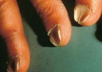
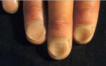
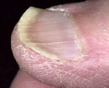
Koilonychia, the opposite of clubbing, is a condition of the person’s nails which has a characteristic of flat or depression of the convexity of the person’s nails. It is most often associated with the disease condition particularly iron deficiency anemia. Some experts would described a person having this disease as someone who have spoon-shaped nails. The nails of persons who are inflicted with this kind of condition, have soft and thin nails. Aside from that, the nails of persons with Koilonychia has a characteristic of brittle and thin nails. Still others would define it as a dystrophy or an atrophic kind of deformity of the person’s fingernails which may be brought about by a lot of factors which will be defined further as we go along. The nails of this persons will take longer time to return to the normal phase. This kind of condition may be either associated with other disease or syndromes or perhaps inherited as this kind of disease is an autosomal dominant kind of disease. The person having this disease condition actually will experience the disorder that affects the nail color, texture, shape and thickness of either the person’s fingernails and toenails. There is a big possibility that the person with koilonychia will have ridges, yellow colored nails and will be susceptible to opportunistic infection. As the condition becomes worse, without treatment, the nails may eventually fall off or perhaps the person will experience a permanent damage of their nails. It is actually a symptom which may be associated by a disease condition.

Koilonychia Causes

Persons with this kind of condition, specifically koilonychia, have acquired the condition because of the following associated causes or etiological factors:

  • Chronic iron deficiency anemia
  • Hereditary forms such as: Adenoma sebaceum, Ectodermal dysplasia, Osteo onychodysplasia also knowned as nail-patella syndrome
  • Sulfur-protein deficiency
  • Trauma
  • Malnutrition
  • Illness
  • Chemotherapy side effect
  • Dysphagia
  • Darier’s disease
  • Excessive usage of oils and soaps
  • Thyroid dysfunction
  • Musculoskeletal condition
  • Renal disease
  • Contact dermatitis
  • Raynaud’s disease
  • Systemic lupus erythematosus
  • Cheilosis
  • Glossitis
  • Pica
  • Hypoplastic patella
  • Acromegaly
  • Hypothyroidism
  • Porphyria
  • Frosbite
  • Thioglycolate depilatory
  • Thermal burns
  • Petroleum based solvents
  • Polycythemia vera
  • Plummer vinson syndrome
  • Infections like yeast or fungal disease
  • Dermatoses like: Psoriasis, Lichen planus, Acanthosis nigricans
  • Carpal tunnel syndrome
  • Hematological conditions like: Hemoglobinopathy, Hemochromatosis
  • Exposure of the person’s nails to nail polish or moisture which lead to brittle nails and peeling of the nail’s edges
  • Nail biting

In determining the cause of the person’s koilonychia, it is adviseable that the person performs diagnostic confirmatory examinations via the doctor’s orders or suggestions to be able to know the exact cause and to be able to pinpoint the treatment for the cause of the disease condition. It is necessary for the person with this condition to approach a trusted and expert physician to allay worries and fear and also to be able to be well informed of why such thing is occuring.

Koilonychia Treatment

With regards to treating the patient with Koilonychia, the treatment will greatly rely on the correcting the underlying cause or deficiency and also being able to manage the problem that is associated with the occurrence of such koilonychia symptom. The only thing that one should bear in mind is that when koilonychia is occuring or is manifested in a person, he or she should undergo treatment as soon as it is recognized to be present to prevent further problems. Aside from that, the following can be done with patients who are positively having the koilonychia such as:

  • Cutting the person’s nails straight and across the top
  • The habit of using softening cream for the skin after washing or bathing
  • Taking iron which is prescribed by the physician and to prevent iron overload, you must follow the prescription
  • Never mix iron supplements with beverages like coffee or tea which is known to stop iron absorption
  • When exposed to harsh chemicals, it is adviced that you wear gloves
  • Consuming vitamins and iron rich foods
  • Wearing shoes that don’t let the person’s toes crowd or squeeze them together
  • Iron supplements must be suggestedly taken 30 minutes before eating
  • Wearing shoes that are comfortable
  • Cautious in exposing or avoid hand exposure especially to chemicals which are harmful

Koilonychia Pictures 

Koilonychia images

Image source – picturesdepot.com

Koilonychia pics

Image source – paraorkut.com

Koilonychia

Image source – nailsmag.com

No responses yet

Posterior Vitreous Detachment – Treatment, Symptoms, Causes

Dec 09 2015 Published by under Diseases & Conditions

Human Eye is a sense organ, small ball in size that allows vision and light perception.

Vitreous humour is a transparent jelly like substances that occupies the space between retina and the lens of the Eye. This clear gel substance provides support to the retina, allows the light to pass through to reach retina and maintains the shape of the eyes posterior chamber. This is made up primarily of water, containing million of collagen fibris supported by hyaluronic acid molecules. Vitreous Humour is connected to retina and fills the space between the lens and the retina of the eye.

As people ages’, Hyaluronic acid begin to decrease and vitreous humour of the eye change and starts to shrink from its original form, this process is called Synchysis. If the synchysis continue to progress, the vitreous humour will separate from the retina and this condition is referred as Posterior Vitreous Detachment (PVD).

About 75% of PVD occur among people over the age of 65, but may also happen for adults under 40s and 50s in age. Research have found that it is more common among women than men.

PVD may happen early if you have eye injury or if you have near-sightedness or Myopia. It has also been reported that people who have PVD in their one eye are likely to have one in the other eye, although for some in might not happen after some years.

Posterior Vitreous Detachment Symptoms

There are situation in which patients with is not aware that they have developed it. Symptoms of PVD is considered as not so serious, but patient will encounter some difficulties:

Floaters

Patients will experience sudden onset of floaters. It can be dot, spots, or wispy lace particles floating across vision. Usually single, but can be also multiple floaters. If multiple floaters is present, it is more suggestive of a Vitreous hemorrhage is associated with Retinal Detachment, much more if positive visual acuity reduction. Patients who experienced Diffuse floaters during eye examinations is more likely suffering from Benign Vitrous syneresis,

Flashes of lights (Photopsia)

These lightning streaks are seen in your peripheral vision. Ocular flashes is experience once the separation of vitreous cause traction over the retina. The retina will get irritated causing it to release electrical impulses. The brain will interpret these impulses as Flashes. It usually lasts for 4 to 12 weeks or may last much longer.

Weiss’ Ring

It is a ring shaped floaters, larger in size as compared to usual floaters that is commonly experience by a person with PVD. It is more clearly seen underlight background. This happen when the vitreous humour was released from the back of the eye.

Posterior Vitreous Detachment Causes

Posterior vitreous detachment is common cause of flashes and floater, though it is not a serious situation, it brings some discomfort to the affected people. Causes of Posterior vitreous detachments are the following:

Age

People over 60 years of age are more common to develop PVD, the risk increases as a person grows older. The vitreous starts to change, and started to reduce its volume that may lead to fall away from retina.

Near sightedness

Near sightedness or popularly known as Myopia, it is a condition that the light produces image in front of the retina. When a persons look in a distant object the visualize object is out of focus, but in focus when looking in a closer object.

Uveitis

Uveitis is the inflammation of the Uvea or the middle layer of the eye. The common cause of Uveitis are autoimmune disorders, infection or it can be exposure to toxins. One of possible complication of Uveitis is Vitreous detachment.

Intraocular laser treatment

This treatment is common to people who are near-sighted, some of them who had received intaocular laser treatment later reports symptoms of PVD.

Intraocular surgery

Most common cause of PVD among younger person is post intraocular surgey. There are about 1% of people who undergone intraocular surgery report incidents of having PVD.

Eye trauma

Patient with injuries in the eyes commonly develops PVD, especially the blunt or any ocular injuries penetrates and cause traction to the vitreous and even to the retina.

Diagnosis

There are various tests available to rule out Posterior vitreous detachment. It is important to have immediate and accurate examination,

not only to confirm the eyes’ condition but also to determine the extent of the said condition. They are as follows:

Dilated eye examination

It is a painless procedure in which the eye is being examined for eye problems like PVD. Using dilation drops, it is instilled in your eyes to widen the pupils. After 30 minutes of instilling dilation drops, the Opthalmologist using equipment will check your dilated eye if there are any problems occurring internally.

Retinal exam

Using a lighted magnifying instruments passing through the retina. Eyes is being scanned, producing digital images of the retina. It can give a clear picture to confirm PVD.

Eye ultrasonography

Ultrasound is a procedure that uses high frequency sound waves that pass through the eye. Reflection of sound waves figure the structure, and measure the size of the eye. There are two types of ultrasonography for the eyes. A-scan ultrasonography is most commonly use to measure eye length conjunction. During cataract surgery it determine the intraocular lens power. The other type is the B-scan ultrasonography is a tool used to evaluate different kinds of eye disorder. It gives the idea on the status of the lens, vitreous, retina, choroid and sclera.

Posterior Vitreous Detachment Treatment

In most cases, Posterior Vitreous Detachment is not sight threatening and does not require treatment since it does not result in permanent loss of vision. Symptoms will gradually subside after a few weeks or months, though people may find it disturbing.

Presence of retinal tear needs further assessment by an Opthalmologist for possible repair to prevent Retinal Detachment to happen.

Surgery

Some Doctors offer treatment to reduce floaters and to prevent it to became bigger. But it is not recognised as a standard treatment in some countries and not practised worldwide.

Exercise

Strenous activity like heavy exercise, running, active sports are avoided six weeks after the appearance of symptoms of PVD. During this period the retina is most at risk for detachment. Follow up check up with your health care provider is recommended after the diagnosis. It will be able to determine if vitreous have completely detached from the retina. Permission from your Doctor to resume on performing heavy activity is a big necessity.

ICD 9 Code

International Statistical Classification of Disease and related health problems or ICD is a medical classification published by the World Health Organization to provide codes to describe diseases and categorized a variety of sign, symptoms andcauses.

No responses yet

© 2017 HealthFoxx.com. All Rights Reserved. Privacy Policy
This website is for informational purposes only and Is not a substitute for medical advice, diagnosis or treatment.