Archive for June, 2014

Estrogen Side Effects, Uses, Advantages

Jun 25 2014 Published by under Side Effects

Estrogen is a group of sex hormones that is used by the body for various processes involving sexual, reproductive and metabolic functions. Estrogen is naturally occurring in the body, but it can also occur as an exogenous drug used for various conditions. It is naturally produced by the ovaries from cholesterol.

Estrogen occurs in the body as estradiol, estrone and estriol. Estradiol is the most predominant estrogen in the body. On the other hand, the other two estrogens are more predominant during pregnancy. Among the three, estrone is the most potent estrogenic substance while estriol is the least potent. Exogenous estrogen is available in various preparations such as:

Uses and Advantages of Estrogen

Estrogen is used for various conditions. General indications of estrogen include:

  • Treatment of hypoestrogenism
  • Hormonal contraception
  • Estrogen replacement therapy in menopausal women and those who have undergone oophorectomy or removal of the ovaries
  • Infertility
  • Management of prostate cancer
  • Relief of symptoms of breast cancer
  • Female hormone maintenance in male-to female transsexuals
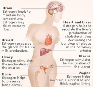
side effects of oestrogen

Estrogen Side effects 

Estrogen plays an important role in various process of the body such as the bones, liver, gene expression, blood clotting, and reproductive functions. The intake of exogenous estrogen may lead to various estrogen side effects related to its oral function. The various estrogen side effects include:

1. Ovarian, Endometrial and Breast Cancer

Estrogen use is also associated with the occurrence of various reproductive cancers as estrogen side effects. This occurs mainly as a result of binding to estrogen receptors that stimulate gene expression, which may damage the DNA and cause cellular proliferation resulting in malignancies.

2. Increase in uterine myomas

The presence of leiomyomas may be increased as estrogen side effect or aggravated by estrogen use because the development of abnormal, benign growths in the uterus is largely dependent on the presence of estrogen.

3. Increased blood clotting

Estrogen also is responsible for the production and utilization of the protein used for blood clotting. Prolonged use of estrogen contributes to the presence of abnormal blood clots leading to thrombophlebitis, deep vein thrombosis, and other clotting disorders that may potentially cause hypertension, stroke, heart attack and pulmonary embolism as estrogen side-effects.

4. Liver diseases

Estrogen use is also linked to the presence of cholestasis or the stasis if bile, which may eventually lead to advanced liver damage as estrogen side effect.

5. Fibrocystic breast changes

Transsexuals and men taking estrogen as hormone replacement or maintenance may have increased size of the breast as estrogen side effect

6. Changes in libido

Those taking estrogen therapies may also suffer from libido changes such as increase or decrease in libido as estrogen side effects. Being a hormone responsible for sexual functioning, initial intake of estrogen may reduce libido because the body is not used to the exogenous estrogen.

7. Nausea and vomiting

Nausea and vomiting are common estrogen side effects. These may occur especially at the start of the therapy and may eventually subside as the body gets used to the exogenous estrogen.

8. Weight changes

Estrogen may also cause weight changes as a result of water retention. People taking estrogen may experience weight gaining as an estrogen side effect.

Aside from the above common estrogen side effects, patients taking estrogen may also experience other estrogen side effects such as dysmenorrhea, scant menstruation, vaginitis, bloating and abdominal cramps, dyspepsia, diarrhea, Erythema multiforme, melasma or chloasma, pruritus, rash, retinal vascular thrombosis, Intolerance to contact lenses, dizziness, migraine, leg cramps, mood disturbances, and hemorrhoids.

In order to prevent these estrogen side effects and other side effects such as Nioxine side effects, proper and prudent use of medications should be employed. In addition, those who have increased risk for the above conditions should not consider taking estrogen. However, when estrogen use is needed, it is important to monitor the estrogen side effects and report them immediately to your health care provider.
No responses yet

Nesidioblastosis – Definition, Symptoms, Causes, Treatment

Jun 22 2014 Published by under Diseases & Conditions

What is Nesidioblastosis?

It is a medical term that means the proliferation of pancreatic islet cell. This often occurs only during the normal development of the neonate. Yet, it can also exist pathologically in infants that will lead to both hypoglycemia and hyperinsulinism. The name was conceived by the person named George F. Laidlaw, who is a renowned scientist, which happened upon the year 1938.  He culled it from the Greek words “nesidia” and “blastos” which means islets and germ, respectively. He described it as a neoformation of the islets found in the Langerhans which is located in the pancreatic duct. He first referred this condition to babies who were born with the pancreatic abnormalities particularly involving the beta cell pancreatic, which are known to produce insulin and is located in the islet of Langerhans of the person’s pancreas. It is an inborn fault of the metabolism part. There is a discordance of the blood insulin and glycemia.

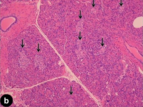
Image of Nesidioblastosis

A histology of the pancreas showing proliferation of cells (shown in arrows)

Others would describe as a medical term that is controversial for the term called hyperinsulemic hypoglycemia episode. It is ascribed to the excess function of the person’s pancreatic beta cells that has an appearance of abnormal microscopic image. In addition to that, this kind of condition is known to be an inherited autosomal recessive disease condition which has, as experts would describe, a characteristic of abnormal formation of the islets found in the person’s pancreas and always having persistent hyperinsulinemia hypoglycemia of the infant person. Others would famously call it as noninsulinoma pancreatogenous hypoglycemic syndrome, congenital hyperinsulinism, islet cell dysmaturation syndrome and nesidiodysplasia. Yet despite of the fact that it most often it occurs reportedly in infants, the adults can’t be spared by this disease condition as they can be also inflicted with nesidioblastosis.

Nesidioblastosis Symptoms

Nesidioblastosis is a dangerous disease condition that affects the person’s pancreas as well as his or her glucose levels. Those who are inflicted with nesidioblastosis will commonly report the following symptoms that they manifest such as:

  • Bloating of the abdominal region
  • Flushing
  • Diarrhea that is watery in consistency
  • Headache
  • Shakiness
  • Blurring of vision
  • Confusion
  • Palpitation of the heart
  • Macrosomic babies
  • Neonatal hypoglycemia that is prolonged

When worse comes to worse, it can also lead to either of the following complications:

  • Damage of the brain
  • Seizure episodes
  • Coma
  • Death

Nesidioblastosis Causes

The cause of nesidioblastosis is due to the following factors such as:

  • Excess of Insulin release
  • Diabetes
  • Defect in the pancreas
  • Abnormal recognition of the glucose
  • Chromosome 11p15.5
  • Acute maternal glucose loading

The cause of nesidioblastosis, which is considered to be a serious kind of medical metabolic disease or disorder, which may be linked to the baby or the adult person as having excess in the release of insulin from the person’s islet of Langerhans which is found in the person’s pancreas.  It may also be associated to obesity and type 2 diabetes.

As mentioned earlier, the person who has nesidioblastosis have an abnormal functioning of the defective beta cells of the pancreas. Other researchers believed that there is a presence of abnormal recognition of glucose because of the defect of the beta cells found in the person’s pancreas. In addition to that, there has been a research that tells that this kind of disease condition is due to genes particularly chromosome 11p15.5.  The combination of the genes for insulin like growth factor as well as the insulin itself is another category that leads to the development of hypoglycemia and hyperinsulinism. Also, when there is a presence of acute maternal glucose loading it may lead to the stimulation of insulin secretion of the fetus which will make them at high risk for having neonatal hyperinsulinism hypoglycemia.

Nesidioblastosis Treatment

Nesidioblastosis can be treated by either:

  • Medical treatment

It is the first thing that is done when a person is diagnosed to have this kind of disease condition. The aim of the medications is to treat the hyperglycemia and hypoglycemia episodes as well as it is also used for the reduction of the symptoms that are seen in persons with nesidioblastosis. The following are the common medications prescribed by physicians:

    • Sandostatin (in either depot IM or Injection)
    • Octreotide Acetate ( either in IM or Injection)
    • Diazoxide
    • Proglycem
  • Surgical treatment

There may be a need for resectioning the pancreas which may be surgically done by a certified surgeon. Other surgeon will suggest doing distal pancreatectomy surgical procedure. Still others will suggest performing subtotal pancreatectomy but there is a risk in this procedure as it may lead to future development of diabetes mellitus or diabetes. Surgical procedure is done when the hypoglycemia episodes is not controlled by medications alone.  The surgical procedure done will be different per individual. It will greatly depend on the progression and the affectation of the disease condition as well as the keen eye of the surgeon.

Clinically speaking, the person who has nesidioblastosis is very difficult to diagnose. There is a need to recognize the disease promptly so as aggressive treatment will be given and be able to avoid any possible and irreversible damage of the central nervous system. With regards to any of the treatment provided to the patient, it is far most important that even after treatment one must be able to monitor the glucose level of the person and be able to at least keep it to a normal level so as to prevent the reoccurrence of such disease condition. This kind of keen monitoring requires skills but the person’s loved ones or the person, himself or herself, can be taught how to do it so as to learn independency. Aside from that, it makes him or her control of at least his or her condition. Also, it is necessary that the person needs to watch not only his or her glucose level but also the foods he or she consumes. He or she should be given the right information with regards to his or her disease condition.

No responses yet

Erysipelas – Pictures, Treatment, Diagnosis, Treatment

Jun 06 2014 Published by under Diseases & Conditions

What is Erysipelas?

Erysipelas is a Greek term that describes reddening of the skin. It is otherwise known as holy fire because of the appearance of skin. It is also termed Saint Anthony’s fire, getting its name from monks who profitably treated the disease. Erysipelas is a superficial infection of the integumentary system, specifically the skin, and caused by streptococcus bacteria. It usually affects the epidermis and extends to the lymphatics. Erysipelas has been present since the Middle Ages and caused the death of several well-known individuals, including Pope Gregory XVI.

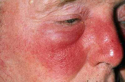
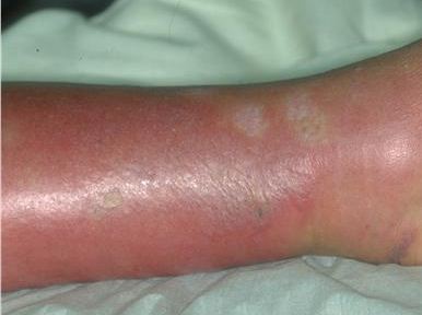
Erysipelas picture 1

Erysipelas Symptoms and Signs

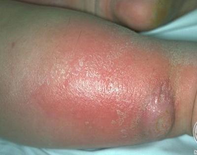
People with erysipelas commonly manifest signs and symptoms such as chills, fever, vomiting, headache, and shaking within two days of infection. Before skin lesions appear, burning and pruritus is common. Skin lesions arise and are characterized as red, warm, swollen, painful and hardened with raised edge or border. The consistency of the lesion or rash is likened to an orange peel.

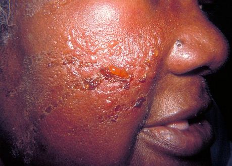
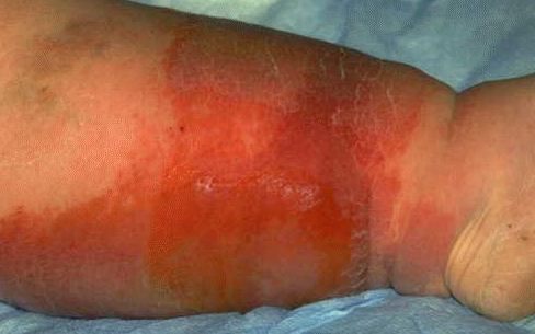
Severe infections result in blisters, bullae, vesicles, petechiae, and necrosis on the skin. The rash spreads and enlarges rapidly in a few hours. It develops on the arms, face, legs, toes and fingers, but the most common site nowadays is the lower legs, accounting for almost 60% of the cases, as well as developing in fatty areas because the microorganism dwells more on these sites. Lymph node swelling may also occur as a result of systemic infection. As opposed to cellulitis, erysipelas does not produce pus. Pus is only seen in complicated and severe lesions. Serous fluids are collected under the lesion and may lead to subcutaneous edema.

Erysipelas Causes and Risk Factors

Causes of Erysipelas

Streptococcus

The most common causative microorganisms are group A beta-hemolytic streptococcus or Streptococcus pyogenes. Other species include non group A streptococcus. These microorganisms produce an exotoxin that leads to rash or lesion on non-infected sites. Streptococci are usually present in the nasal passages and cause illness after spreading to the skin.

Any skin openings

The streptococcus bacteria enter the skin through wounds, abrasions, or any minor trauma.

Risk Factors for Erysipelas

Common risk factors for the disease are:

Extremes of ages

Infants, children, and the elderly are more predisposed to develop the infection because of a compromised immune system.

Co morbidity or any underlying disease

Presence of diabetes mellitus, fungal infections, skin ulcerations, and infections increase the risk of developing erysipelas because of poor wound healing and entrance of streptococcus bacteria.

Impaired lymphatic drainage

Cases such as pelvic surgery, mastectomy, and grafting impair lymphatic circulation thus increasing the risk of developing infections.

Immune deficiency states

Decreased immune systems in cases of steroid therapy, malignancy, chemotherapy and other diseases predispose a person to acquire an infection such as erysipelas.

Erysipelas picture 2


Erysipelas Diagnosis

Diagnosis of the disease and differentiating it from other skin infections relies on the appearance of the rash, well-demarcated and raised. An antistreptolysin O titer may be determined through blood tests after eight to ten days of infection to determine infection by a streptococcus.

Erysipelas Treatment

General symptoms of the disease generally disappear in three days while skin abnormalities usually return to normal in several weeks. Treatment of the infection involves administration of antibiotics and supportive management.

Prompt medical consultation

Erysipelas is a serious skin infection because it causes several complications involving major organs such as the heart and kidneys. Erysipelas do not heal on their own as compared to other self-limiting diseases, hence it requires prompt diagnosis and effective medical treatment.

Antibiotic therapy

Oral and intravenous antibiotics are administered to patients to destroy the bacteria. The most common antibiotic class used is penicillin and is administered for one to three weeks. Patients allergic to penicillin may be given alternative medication such as cephalosporins and macrolides. Mild forms involve administration orally but more advanced cases require intravenous antibiotic treatments. Infection from staphylococcus aureus should also be considered, hence a penicillinase-resistant antibiotic should also be used. Hospitalization for these cases is required for continuous monitoring and IV medications. To prevent recurrence of the infection, prophylactic antibiotics are also given.

Wound Care

Prevention of re-infection and complications include proper wound care. Use antiseptic solutions and creams to properly clean the area.

Dressing application

Dressings may be applied as long as it is changed every day to prevent further infection. Wet dressings are considered for necrotic lesions and should be changed two to three times a day.

Debridement

Patients who develop gangrene and necrosis may need debridement to allow faster tissue healing. Debridement involves the removal of dead skin or tissues to make way for newer ones.

Elevation of the extremities

Swelling and edema is greatly reduced by elevating the affected extremity to enhance venous and lymphatic flow. Elevate the body part with two to three pillows so as not to compromise arterial blood flow.

Use of compression stockings

These devices improve venous blood return and reduce swelling, especially in patients with impaired lymphatic drainage.

Erysipelas Prognosis

There is an excellent prognosis of the disease and takes about several weeks to heal and disappear. The infection is well-treated by the use of antibiotics. Deaths which occurred during early times were a result of an absence of specific treatment for the infection and advanced complications. Today, large spectrum antibiotics are present to effectively eradicate the bacteria.

Erysipelas Complications

Streptococcus bacteria may spread to other organs and give rise to the following complications:

Abscess and gangrene

Further tissue damage on the lesion may lead to pus formation and even death of the tissue.

Glomerulonephritis

Group A beta hemolytic streptococcus commonly infects the kidneys and produces inflammation on the glomerulus, leading to a decrease in urine output and water retention.

Thrombophlebitis

Serous fluid that collects under the skin may lead to thrombus formation and inflammation of the veins.

Endocarditis

As with glumorulonephritis, streptococci also travel to the heart and infect the endocardium (the inner muscle layer of the heart).

Toxic Shock Syndrome and Sepsis

Advanced infection allows the microorganism to replicate in the blood stream and subsequently impairs the blood vessels, leading to hypotension.

Joint and Bone diseases

Arthritis and bursitis may occur as a result of joint infection and synovial fluids.

Reoccurrence of the infection

Ten to twenty percent of patients experience re-occurrence of infection even after antibiotic therapy.

Lymphatic infection

Microorganisms and debris drains into the lymphatic system where it can proliferate again.

Necrotizing fasciitis

It is otherwise known as the “flesh-eating disease” because it involves a deeper layer of tissues. Almost 80% of patients experiencing this complication have poor prognoses.

Erysipelas Prevention

Prevention of erysipelas involves prevention of wound infection and prompt treatment of any open wounds. Good personal hygiene is also essential to prevent harboring of microorganisms such as streptococcus. Increasing immunity such as increasing vitamin and mineral intake and a balanced diet with appropriate exercise is also helpful in preventative care. The most important prevention is to avoid skin breakage because it is the primary portal of entry for streptococcus. Care for the skin by hydrating and protecting it from bio-mechanical trauma.

Erysipelas Pictures

Erysipelas

Erysipelas Pictures

Erysipelas Images

Erysipelas image

No responses yet

Dermatofibrosarcoma Protuberans – Pictures, Symptoms, Treatment

Jun 01 2014 Published by under Diseases & Conditions

What is Dermatofibrosarcoma Protuberans?

Dermatofibrosarcoma Protuberans is a rare soft tissue growth that is slow-growing, not often metastasize (less than 4% of total number of cases), in some cases of spreading, the lungs are the most common direction. It frequently recurs or remains. It can enter both the muscle and bone. It happens in all ages, but most commonly among adults, from early on up to middle adulthood. The average age of onset is 40 years. It is one of the rarest among all skin cancers; approximately less than one per 100,00 cases are recorded every year. It is the most common cutaneous sarcoma.

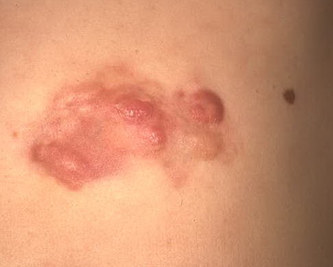
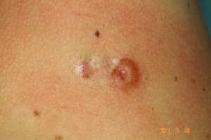
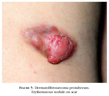
Dermatofibrosarcoma protuberans pictures

Symptoms of Dermatofibrosarcoma Protuberans

Tumor

Tumors usually start as slow-growing. The progression is also delayed. It usually starts in a small papule or small patch that appears anywhere in the body, but frequently on the trunk. Tumors are usually smooth and even, sclerotic and yellowish in color. The tumor grows in a particular direction, extends upward and downward, following the path of the least resistance. It later develops in a larger nodules.

Variants:

  • Bednar Tumor

A pigmented form of DFSP occurring predominantly in African descendants and representing less than 5% of all DFSP. It has a lower recurrence rate.

  • Myxoid DFSP

Characterized by the interstitial accumulation of hyaluronic acid ground substance, rendering the usual storiform pattern less pronounced and a nonpatterned array of vessel more apparent.

  • Giant cell fibroblastoma

A rare mesenchymal tumor occurs predominantly in the first decade of life, showing an unidue combination of spindle cell patterns, myxoid aread, pleomorphic and multinucleated giant cells, and distinctive sinusoid-like spaces. Local recurrence is common but metastasize is not

Stages:

Stage 1

Primary tumor appears as well as localized disease

Stage 2

Metastasize occurs in the Lymph nodes

Stage 3

Distal Metastasize happens

Diagnosis of Dermatofibrosarcoma Protuberans

Histologic Findings

The characteristic of Dermafibrosarcoma protuberans under microscopy is a widely-spreading dermis and subcutenous tissues. The tumor can get through the space between the subcutaneous tissue. Aspiration using fine needle smears are commonly hypercellular and stroma poor. The mitotic activity is usually low; inflammatory infitrates, foamy histiocytes, multinucleated cells and hemosiderin deposits are not common. The overlying epidermis is generally thin or ulcerated.

According to Layfield, the following results are observed in Dermafibrosarcoma:

a. Smears of moderate cellularity containing single cells and cells entrapped within tissue fragments, stromal fragments with a fibrillar character

b. Plump oval to spindle isolated cells with bland nuclear features and a fine chromatine pattern

c. A moderate amount of cytoplasm with a pale, granular appearance

d. Rare mitotic figures

Differential Diagnosis

The following should be made different with Dermafibrosarcoma protuberans.

a. Low grade spindle sarcoma

b. Benign fibrous histiocytoma

c. Desmoids

d. Low grade malignant peripheral nerve sheath tumor

Other diagnostic procedures include:

Chest Xray

Since the lungs are the common site of metastasize, screening for pulmonary spread out is very important.

Computed Tomography Scanning

To detect if there is bone or muscle involvement.

Magnetic resonance Imaging

To define the periphery and the extent of the tumor.

Fluorodeoxyglucose (FDG) Scanning

To monitor the degree of metastasize.

Treatment of Dermatofibrosarcoma Protuberans

a. Surgery

Surgical removal of the tumor is the primary and best treatment for Dermatofibrosarcoma protuberans. It is called Mohs Surgery; it takes away not only the tumor but the pathological cells also. The rate of Mohs surgery is highly successful.

b. Chemotherapy

Use of oral drugs like Imatinib produce positive results to reduce the tumor in patients with Dermatofibrosarcoma protuberans

c. Radiation Therapy

Radiation Therapy is more present compared to the past. It is recommended to prevent deficits in the functional or appearance of a person. It also lessens the risk of recurrence; its risk of complication is also low.

Prognosis

The prognosis for Dermatofibrosarcoma protuberans has a higher chance of survival if detected early. Malignancy is rare and the incident of Metastasis is uncommon. But in some cases, the tumor recurs. It has an average of approximately 50% chance of coming back. Tumors can result in death if they become malignant.

ICD9 code

ICD-9 Codes

173.90 – Unspecified malignant neoplasm of skin, site unspecified

Dermatofibrosarcoma Protuberans Pictures

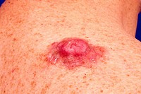
Dermatofibrosarcoma protuberans papule photo

A starting tumor at the trunk

Dermatofibrosarcoma protuberans picture

Picture of Dermatofibrosarcoma protuberans tumor

A single erythematous tumor with small surrounding papules

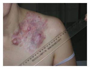
Several Tumors in Dermatofibrosarcoma protuberans pics

Diffused Dermatofibrosarcoma protuberans at the chest area

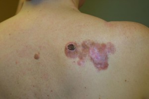
Advanced Dermatofibrosarcoma protuberans image

Necrotic Dermatofibrosarcoma protuberans showing a dark spot on the back

No responses yet

© 2017 HealthFoxx.com. All Rights Reserved. Privacy Policy
This website is for informational purposes only and Is not a substitute for medical advice, diagnosis or treatment.