Archive for September, 2014

Groin Hernia – Pictures, Symptoms, Surgery, Recovery, Repair

Sep 26 2014 Published by under Diseases & Conditions

What is Groin Hernia?

Groin hernia, according to statistics occurs to about 4.4 percent in children and about 98 percent in children that ages 13 years old and below. Most of the children that are inflicted with groin hernia have indirect form of groin hernia which is approximately 99 percent than that of direct groin hernia which accounts for about 1 percent only.

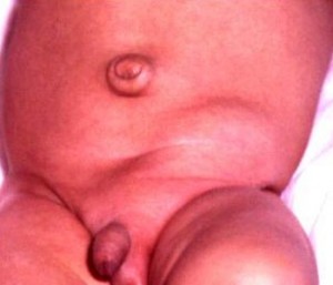
Groin hernia is actually a bulge found in the external portion of the person’s groin. It occurs when there is a spot that is weak which is found in the wall of the abdominal area. It is known to contain connective tissue, intestinal portion as well as fat. There are actually two types of groin hernia which are femoral and inguinal hernia. The groin hernia is interchanged with the inguinal hernia which accounts mostly common hernia that happens in adults.

Groin Hernia picture

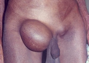
Groin hernia showing protrusion in the groin

Groin Hernia Symptoms

  1. Groin Hernia in Men

According to research and statistics, groin hernia occurs mostly on men than on women. Approximately in America alone, 500,000 men or more each year undergoes surgical repair of their hernia. Groin hernia in men occurs when the intestinal loop enters in the person’s inguinal canal which is located in the groin area which anatomically speaking is located just in between the person’s pubis and the leg’s top portion. It occurs most commonly in men who are known to do the heavy works such as picking up objects that are heavy without the use of proper body mechanics. The common groin hernia type that occurs most in men are inguinal hernias.

  1. Groin Hernia in Women

The type of groin hernia that occurs in women is femoral hernias. This kind of groin hernia happens to women because anatomically speaking the women is known to have a bone structure that is wider compared to the men. The femoral hernia, which is a type of groin hernia, is not that commonly reported. According to statistics, it occurs for about 3 percent than all other hernias combined. There are five kinds of groin hernia that occurs in women such as:

    • Strangulated groin hernia in women

This occurs when the groin hernia will produce a blockage of the supply of blood towards the bowel part.  This kind of hernia is a medical emergency kind. This may also occur in men.

    • Obstructed groin hernia in women

It happens when an intestinal part is intertwined with hernia leading to the obstruction in the intestinal portion. This may grow and may be very painful as it progress. In worst case scenario, vomiting can happen.

    • Irreducible groin hernia in women

This occurs when the hernia, particularly the femoral hernia, is stuck in the femoral canal which results in the experience of pain and illness feeling.

    • Reducible groin hernia in women

It occurs when the hernia can be push normally back into the person’s abdomen either via manipulation or done spontaneously. This kind of groin hernia is a painless kind.

    • Incarcerated groin hernia in women

Incarcerated groin hernia is a medical term that means obstructed form of hernia that can be irreducible but not really strangulated kind.

Groin Hernia Causes

When a person has tears or weakness in his or her abdominal wall it can lead to the person to have groin hernia which may be due to the following etiological reasons such as:

  • Previous surgical procedure done
  • Weakness of the abdominal wall which is age related
  • Defects during birth
  • Wear and tear that is prolonged and situational such as coughing or straining that is lifting

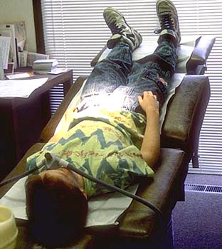
Groin Hernia Diagnosis

In diagnosing groin hernia, the physician usually does the following diagnostic tests such as:

  • Physical examination
  • Medical historical examination
  • Ultrasound examination
  • Computed Tomography scan examination
  • X-ray examination

Groin Hernia Treatment

Usually, groin hernia will not require treatment. Yet, those persons with groin hernia that leads to the production of symptoms that leads to the enlargement of the groin hernia itself needs to be corrected or repaired by a licensed surgeon. The following treatments are done to persons with groin hernia:

  • Supportive device

Prior to the surgical procedure, the person with groin hernia are expected to use truss, which is a supportive device that is worn to provide pressure on the hernia area and keeps control of the hernia itself.

  • Non surgical treatment

Non surgical treatment can be another form of treating persons with groin hernia such as:

    • Weight loss
    • Sleeping with the upper most pat of the body being propped up
    • Don’t eat before lying to bed
    • Stop smoking
    • Eating fiber rich foods
    • Being able to properly lift things
  • Surgical procedure

The effective and safest way of treating groin hernia is done via surgical procedure. The type of surgical procedure done will be made via the assessment of the physician in charge. It may be done via open approach, herniorrhaphy, bowel resection, hernioplasty or via laparoscopic approach.

Groin Hernia Repair

Groin hernia repair is done via surgery. The following surgical repairs commonly done by surgeons are as follows:

  • Laparoscopic surgical repair

In this procedure, the surgeon usually does small incisions in the person’s abdomen and will then inflate the abdomen via a gas that is harmless in nature. After which, the laparoscope is inserted. A laparoscope is an instrument that consists of surgical instruments and minute video camera. The long term rate of success with this surgical repair procedure is less compared to that of the open surgical repair. The advantage of this surgical repair procedure is that it heals quickly with lesser pain associated.

  • Open surgical repair

This kind of surgical repair is done via local or general anesthesia. What happens in this surgical repair is that an incision is made in the person’s groin and a small synthetic mesh material reinforces the area of groin hernia is used to prevent hernia from coming back or producing another one.

Groin Hernia Recovery

Hernia in groin which is usually treated surgically, will recover around a week or two. Hence, certain things should be done to ensure the recovery of the person who was surgically treated from inguinal hernia. The following things should be considered under groin hernia recovery:

  • Healthy diet which is high in fiber, rich in vegetables and fruits
  • Prevention of lifting heavy things for at least a month or so
  • Pain relievers are taken for pain episodes
  • Rest periods are provided

By doing such things, recovery for persons with hernia in groin will be achieved.

Groin Hernia Pictures

Hernia Support picture

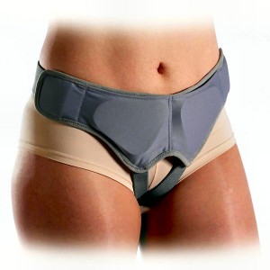
Supportive device used to reduce pressure on the hernia

Groin Hernia image

Inguinal hernia in an infant

Groin Hernia picture

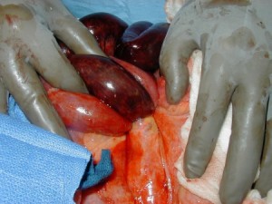
Strangulated hernia showing gangrenous bowels
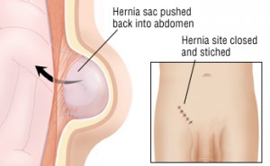
Groin Hernia repair

Hernia repair

No responses yet

Inclusion Body Myositis – Prognosis, Symptoms, Treatment, Causes

Sep 20 2014 Published by under Diseases & Conditions

What is Inclusion Body Myositis?

Inclusion Body Myositis or IBM is what is medically termed as an inflammatory muscular disease. It is characterized as a progressive muscle wasting and weakness. Persons aging 50 years or more are the people who are often affected by this disease condition. These are a very rare condition that occurs and is reportedly seen most on women than in men. Although this said muscular disease condition can be seen during adulthood.

Inclusion Body Myositis (IBM)

Observe Muscle wasting of both thighs

Causes of IBM

The exact etiology of Inclusion Body Myositis is still unknown. Yet there are theories with regards to the causative factors, such as:

Autoimmune disease

Researchers theorized that it is a form of autoimmune disorder wherein the immune system is in constant fight of the infection until it disappears. Hence, by mere fighting of the immune system, aside from it destroys the virus; it also destroys healthy tissues and putting a bigger risk for the person’s health. It is the mere fact of the patient’s body attacking its own muscular tissues causing swelling and inflammation within the person’s muscular system.

Degenerative disease

Another theory is that it is a degenerative kind of disease wherein the degeneration or atrophy and aging process of the person’s muscles leads to an abnormal buildup and damage of protein.

Medication

Another theory suggested that this disease condition called Inclusion Body Myositis or sometimes called as Sporadic Inclusion Body Myositis is due to the fact that the person takes medication that his or her body is allergic to. Aside from that certain medications made available to the public can cause Inclusion Body Myositis. Some of these medications include cholesterol- lowering drugs, penicillamine, carticaine, interferon-alpha, growth hormones, carbimazole, cimetidine, phenytoin, hepatitis B vaccine. The medications mentioned all contribute to developing Inclusion Body Myositis. It is only through stopping the intake of such medications that the symptoms are relieved.

Genetics

It is believed that one of the causative factors that predispose a person to the said disease condition is the fact that he or she has the gene mutations in the UDP-N-acetylglucosamine 2-epimerase/N-acetylannosamine kinase (GNE) gene. With this gene mutation, it is theorized that the gene impairs the function of the GNE protein leading to having this kind of muscular disease condition.

Infections, Injuries and Viruses

These are other causative factors that contribute to the Inclusion Body Myositis. Acquiring an injury into the patient’s muscles, such as a sport incident or a car accident, may lead to inflammation and edema of the muscular tissue. Viruses, on the other hand, trigger the autoimmune Inclusion Body Myositis symptoms. Persons who are HIV positive can lead to the development of this muscular disease condition.

Symptoms of IBM

When a person has been diagnosed with having Inclusion Body Myositis, he or she will manifest the following symptoms:

  1. Muscle weakness that slowly progresses in a matter of months or years
  2. History of Frequent falls
  3. Dysphagia or having difficulty swallowing
  4. Limb weakness
  5. Head drop or difficulty holding the head up
  6. Muscle wasting
  7. Mild facial weakness
  8. Increase in difficulty in the day to day task that requires the usage of the muscles located in the proximal area, such of these tasks includes combing the hair, lifting objects, climbing steps, getting up from a chair and stepping onto the curb.
  9. Poor grip
  10. Pain or discomfort as muscles weaken
  11. Weakness is asymmetrical
  12. Diminishing DTR or deep tendon reflexes
  13. Mixed neurogenic and myopathic changes on electromyography
  14. Weakness in the patient’s fingers, wrist, thighs and forearms.

The symptoms mentioned may vary from person to person.

Diagnosis of IBM

The person is thoroughly physically examined by a physician along with questioning related questions with the disease condition and also letting the person take test to confirm the diagnosis. Such of these tests includes the following:

  1. Muscle biopsy
  2. Blood Test
  3. MRIs or magnetic resonance imaging
  4. EMGs or electromyography
  5. Nerve conduction studies
  6. Lab results of Serum creatinine kinase which is less than 12 times the normal

Additional laboratory values that are considered elevated with this kind of disease condition includes:

  1. Serum aldolase
  2. Serum lactate dehydrogenase
  3. Serum alanine aminotransferase
  4. Serum aspartate aminotransferase

Aside from that to be diagnosed with this kind of disease condition, one must possess the following clinical features or criteria:

  1. Onset age of greater than thirty years old
  2. Illness duration which is greater than six months
  3. And some of the symptoms that were mentioned above.

It is often misdiagnosed as polymyositis and the person is diagnosed with this one if the patient doesn’t respond to the therapy intended for polymyositis.

Treatment of IBM

When a person has this disease condition, he or she together with his or her physician will work towards the goal of the treatment. The treatment goal focuses on the muscle strength improvement in order to improve the activities of the daily living or ADLs. One must also know that when one is diagnosed to having this muscular disease condition, there will be no treatment that can slow or stop the disease progression.

Immunosuppressants. The specific therapy for this kind of disease condition includes immunosuppressive therapy or treatment.

Intravenous globulin or steroids. Corticosteroids or steroids for that matter help in the regulation of the immune and inflammatory response.

There is also a need for multidisciplinary treatment approach for:

  1. Speech and language therapy in the dysphagia assessment
  2. Physiotherapy and occupational therapy
  3. Dietary support since the patient is at risk for choking due to dysphagia

Prognosis of IBM

According to studies, when one is diagnosed with having Inclusion Body Myositis, there is actually a slow progression when this kind of muscular disease condition is manifested in adulthood. However, if the symptoms are manifested in old age, the muscular disease condition tends to progress in a rapid manner. When one has progressive dysphagia, there is a poor prognosis of the patient.

No responses yet

Fentanyl Side Effects

Sep 16 2014 Published by under Side Effects

Fentanyl is a narcotic analgesic, which has a short duration of action and rapid onset. Fentanyl is manufactured in the brand names of Matrifen, Lazanda, Abstral, Instanyl, Onsolis, Haldid, Fetora, Duragesic, Durogesic and Actig. It is an agonist of the opioid receptors, which significantly limits the transmission of pain impulses by decreasing the calcium electrolyte levels inside the cells.

fentanyl side effects

Uses of Fentanyl

Fentanyl was historically used for the treatment of breakthrough pain. It is given in patients before surgical procedures in conjunction to benzopdiazepines as an anesthetic. The vast uses of fentanyl in pain relief made way for the manufacture of fentanyl citrate, which is widely used as a general anesthetic agent. There were additional fentanyl analogues, which have been developed. Fentanyl also became a palliative treatment in the form of Duragesic patch, Fentora buccal tablets and Actig lollipop. Historically, fentanyl was also used to incapacitate and to kill in military applications. Because of its effects, it has also been used as a recreational drug, causing fatalities of several people. Today, the approved uses of Fentanyl include:

  • Anesthetics for surgical procedures such as in epidural or intrathecal routes
  • Analgesics in patients in intensive care units needing pain relief
  • Sedation for cardiac catheterization, endoscopy and oral surgery in conjunction with midazolam, a benzodiazepine drug
  • Management of chronic pains such as the presence of cancer pain
  • Adjunct anesthetic for root canal treatment and pulpectomy
  • Pain relief for patients with renal failure
  • Pain relief for patients that cannot tolerate other forms of opioids such as morphine

Fentanyl Side effects

The side effects of fentanyl are vast and it may affect all the organ systems in the body. Careful use of the drug will usually not result to these fentanyl side-effects. Fentanyl side effects include:

1. Nervous system Fentanyl Side effects

Mental Depression

This fentanyl side effect is rooted on the inhibition of nerve impulses in the brain. Patients may suffer from delirium, stupor, dysphoria and somnolence similar to carbamazepine side effects.

Myoclonic Seizures

This type of seizure has also been observed in patients using transdermal therapy. These seizures may have occurred due to fentanyl induced rigidity.

Migraine

A case of hemiplegic migraine has also been documented with the use of fentanyl for sedation.

Drug abuse

Fentanyl has potential for drug abuse because of development of drug dependence.

Withdrawal

Stopping fentanyl may lead to withdrawal syndrome because of possible drug dependence.

2. Cardiovascular Fentanyl Side effects

Hypotension

Fentanyl may also cause hypotension because of possible vasodilating effects. Safety should be ensured to prevent falls.

Bradycardia

Fentanyl may aldo cause slowing of the heart rate due to CNS depression and possibly slowing down of the action potentials in the heart as well.

3. Gastrointestinal Fentanyl Side effects

Nausea and Vomiting

The opioid receptors in the gastrointestinal system may also be affected leading to nausea and vomiting because of slowed peristalsis of the GIT.

Constipation

Slowed peristalsis may also lead to constipation. Increased fiber and fluid intake may reduce this fentanyl side effect.

Choledocoduodenal sphincter spasm

Rare occurrence of spasm in the sphincter of the common bile duct in the duodenum has been documented. This may be due to fentanyl induced rigidity.

4. Respiratory Fentanyl Side effects

Respiratory Depression

The inhibition of nerve impulses in the central nervous system also slows the action of the respiratory center of the brain, which is located on the brain stem. Probably, this is the most serious fentanyl side effect because it can bring about respiratory failure leading to death. Observe for shortness of breath, decreased rate of breathing and shallow respirations.

5. Genitourinary Fentanyl Side effects

Urinary retention

The urinary system also has opiate receptors leading to urinary retention when fentanyl binds with its receptors in the bladder.

6. Dermatologic Fentanyl Side effects

Pruritus and rashes

Rashes and pruritus have been documented due to mild hypersensitivity reactions especially in the use of transdermal patches.

7. Hematologic Fentanyl Side effects

Hemolysis

Destruction of RBCs was seen in patients receiving large volumes of hypotonic fentanyl via the intravenous route. In order to prevent this, injections should be in slower rates or use of isotonic solutions.

8. Metabolic Fentanyl Side effects

SIADH

Syndrome of inappropriate anti-diuretic hormone involves the presence of high amounts of anti-diuretic hormone, leading to water retention and edema. This fentanyl side effect is rarely developing.

9. Immunologic Fentanyl Side effects

Increased Killer cells and CD 16+ lymphocytes

The levels of these white blood cells have been increased in some patients receiving fentanyl although the exact mechanism is still unclear.

Since fentanyl side effects are vast, physicians prescribing it usually use caution in dosing because over dosage increases the risk for life-threatening fentanyl side effects such as respiratory depression. Despite thesefentanyl side effects, it is still beneficial for patients requiring pain relief and bedsides; it is considered the safest opioid analgesic, despite its fentanyl side-effects.

No responses yet

Peripheral Artery Disease – Symptoms, Treatment, ICD 9, Causes

Sep 10 2014 Published by under Diseases & Conditions

What is Peripheral Artery Disease?

Peripheral artery disease (PAD) is a condition arising from the obstruction of the large arteries in the peripheral areas of the body such as the hands and legs. Peripheral artery disease is otherwise known as peripheral vascular disease (PVD) or peripheral artery occlusive disease (PAOD).

Obstruction in the arterial wall may result from inflammatory processes and atherosclerosis on the arteries causing thrombus, embolus or stenosis formation. The obstruction in the arteries often leads to ischemia.

Peripheral artery disease also includes micro vascular disorders involving constriction of the arteries such as Reynaud’s syndrome or dilation of the arteries such as erythromelalgia.

PAD affects up to 14% of the general population mainly affecting those ages 70 years old and above. Most of the cases are also asymptomatic and do not require extensive treatment and amputation. In fact, only 25% of the patients undergo treatment. Three out of 10 diabetics also suffer from PAD.

Peripheral Artery Disease 1

The obstruction associated with PAD also increases the risk of developing heart attack and stroke.

Peripheral artery disease is divided into four stages as introduced by Rene Fontaine last 1954:

  1. Incomplete blood vessel obstruction, mild pain during walking (claudication).
  2. Severe pain during ambulation involving only short gaps or distances (referred to as intermittent claudication)
  3. Stage IIa- after walking a gap of greater than 150 meters
  4. Stage IIb- after walking a gap of less than 150 meters
  5. Pain during resting, aggravated by raising the limbs (rest pain)
  6. Tissue loss and difficulty in ambulation (gangrene)

Rutherford introduced a more advanced categories including six types:

  1. Mild pain
  2. Moderate pain
  3. Severe pain
  4. Ischemic pain even when not involved in any activities
  5. Slight tissue damage
  6. Gangrene

Pathogenesis of Peripheral Artery Disease

Presence of any injury to the lining of the arteries causes inflammation to the area. This process results to clot formation and platelet aggregation forming a thrombus or plaque to heal the damaged area. Once this thrombus increases in size, it significantly reduces blood flow to the proximal areas of the clot. Stenosis also results, which is the narrowing of the lumen of the arteries as a result of atherosclerosis (build-up of fats). Reduction in blood flow causes reduced oxygenation leading to ischemia. The tissues undergo anaerobic metabolism releasing lactic acid from fats and proteins. The build-up of lactic acid causes pain and claudication. Chronic ischemia leads to death of the tissue or gangrene that needs amputation.

Furthermore, a thrombus can be dislodged and become an embolus. When this happens, the clot formation can lodge to small arteries in the heart or brain, leading to heart attack and stroke.

Causes and Risk factors

Risk factors of peripheral artery disease include:

Old age

The incidence of PAD increases as the age increases. People aged 70 years old and above are of greater risk for PAD because of reduced elasticity of the blood vessels.

Gender

Men are more prone to developing PAD because of certain habits such as smoking.

Smoking

Smoking is one of the major causes of PAD and increases the risk by tenfold. Nicotine in cigarettes is a potential vasoconstrictor which reduces the lumen of the arteries.

Hypertension

High blood pressure creates injury to the arterial walls leading to platelet aggregation and clot formation. Hypertension is also directly associated with cardiovascular and cerebrovascular accidents.

Hyperlipidemia

High levels of low-density lipoproteins (“bad cholesterol”) speeds up atherosclerosis which significantly blocks the arteries.

Diabetes Mellitus

Patients with diabetes also have increased risk for atherosclerosis formation due to sluggish blood flow brought about by high blood glucose levels. Most cases of leg amputations are associated with patients who are diabetic.

Signs and Symptoms of Peripheral Artery Disease

Most of the patients with PAD are asymptomatic. Other symptoms include:

Caludication

Claudication refers to pain, numbness, cramping and weakness of the extremities because of poor blood flow.

Wounds that heal slowly

Poor blood circulation to the injured area reduces the transport of white blood cells, platelets and clotting factors that help heal the injured part.

  1. Cold, clammy skin on the affected part
  2. Paleness or cyanosis of the extremities
  3. Diminished nail and hair growth over the affected extremity
  4. Weak and thready pulse in the affected limb, some serious obstructions may lead to absent peripheral pulse
  5. Erectile dysfunction

Patients with PAD and diabetes may experience the symptom.

Diagnosis

Diagnostic test aims on determining any obstruction of blood flow in the peripheral areas of the body. Certain blood tests are also used to confirm presence of underlying factors such as hyperlipidemia. The tests include:

Ankle brachial pressure index

This test involves measuring the blood pressure in the arms and legs (particularly in the ankles). When the pressure of blood in the legs is lower than the pressure in the arms, PAD is suspected. Arterial brachial index ratio is also computed. ABI ratio of less than 0.9 is definitive with PAD. An ABI less than 0.8 indicate average disease and an ABI ratio of 0.4 is used as a baseline.

Doppler Ultrasound

Doppler ultrasound examines the blood flow to the limbs through the use of sound waves. A handheld device is placed over the affected areas. Sound waves are then converted to pictures of blood flow to determine any obstruction.

Treadmill test

ABI tests are done before and after the patient use treadmill. Significant changes in the ABI ratio during these times indicate severity of symptoms during a particular level of activity or exercise.

Arteriogram

Arteriogram involves the injection of a dye in the arteries and x-ray imagings are taken. This test determines any obstruction and the location as the dye passes through the arteries. Patients should be assessed for presence of allergies to seafood because the dye is composed of iodine based material.

MRI

Magnetic resonance imaging is one of the most accurate tests for PAD to locate the obstruction and describe the severity.

Blood tests

Cholesterol levels, triglycerides and blood glucose levels are determined to rule out any underlying conditions such as diabetes and hyperlipidemia.

Treatment of Peripheral Artery Disease

Treatment of PAD involves certain medication and lifestyle modification

Medications

Anti-platelet aggregate medications

Aspirin and clopidogrel are antiplatelet medications that reduce platelet aggregation to reduce thrombus formation. Ensure safety to prevent bleeding episodes and watch out for signs of bleeding in the body.

Antihyperlipidemics

Statin drugs such as simvastatin and atorvastatin inhibits the synthesis of lipid in the body.

Lifestyle Modification

Exercise

Exercise improves circulation and significantly reduces claudication. Treadmill or brisk walking is beneficial for PAD involving the lower extremities.

Smoking cessation

Cessation of smoking reduces the incidence of PAD and relieves symptoms

Lower blood pressure by means of diet and compliance to antihypertensive medications.

Surgery

Angioplasty

This involves the insertion of catheter balloon into the sclerotic artery to widen the lumen of the area.

Plaque excision or Arthrectomy

Plaque excision involves the scraping off of plaque inside the large arteries.

Bypass grafting

Grafting is done in areas of severe stenosis. The saphenous vein in the thighs is usually used to replace the stenosed artery. Artificial materials such as the Gore-Tez may also be used.

Sympathectomy

Sympathectomy is rarely done and involves removal of the nerves that innervate the area that cause vasoconstriction.

Amputation

Removal of the gangrenous area is the last resort to prevent further complications such as renal disease.

Complications

Conditions may arise as a result of continuous impairment in the circulation and embolus formation. Complications of PAD include:

  1. Heart attacks and stoke
  2. Severe ischemia leading to gangrene
  3. Amputations of the gangrenous area

Complications may be prevented by instituting appropriate management and adhering with the suggestions and treatment.

Prevention

Prevention of PAD involves avoiding the modifiable risk factors such as:

  1. Quit Smoking.
  2. Avoid high fat content foods.
  3. Ensure adequate daily exercise.

ICD 9 Code of Peripheral Artery Disease

The ICD or International Classification of Diseases is used to classify and code mortality data from a specific disease. The ICD code for PAD is 443. Specific codes for specific types of PAD include:

443 Other peripheral vascular disease
443.0 Raynaud’s syndrome
443.1 Thromboangiitis obliterans
443.9 Peripheral vascular disease, unspecified

No responses yet

Decompression Sickness – Symptoms, Treatment, Definition, Causes

Sep 05 2014 Published by under Diseases & Conditions

Decompression Sickness Definition

Decompression Sickness is a medical term that commonly affects deep sea or ocean divers but it can also happen to pilots in an unpressurized aircraft. The Decompression Sickness is otherwise known as caisson disease or the bends or primary the diver’s disease or generalized barotrauma disease condition. This kind of disease condition is a combination of the high altitude, aerospace related and changing of the barometric pressure. It is experienced by most divers who encounter a dissolution of the gases that comes out of the solution into the bubbles inside the depressurization of one’s body. It is manifested by divers who undergo rapid decompression or when the divers ascend to the surface in a quick manner will lead to decompression sickness episodes.

Decompression Sickness Symptoms

Persons who are suffering from decompression sickness commonly report the following symptoms:

  • Chest pain
  • Irritation in the skin
  • Neurological problems
  • Pain in the joints
  • Itching
  • Cramps
  • Formication episodes or sensation wherein tiny insects crawls in one’s skin
  • Skin swelling
  • Pitting edema
  • Abnormalities in the vision
  • Seizure
  • Behavior changes
  • Fecal incontinence
  • Urinary incontinence
  • Fatigue that is unexplained
  • Skin rashes
  • Headaches
  • Vertigo
  • Nausea
  • Loss of hearing
  • Loss of balance
  • Vomiting
  • Malaise that is generalized
  • Paralysis
  • Confusion
  • Cough that has a presence of blood and frothy kind of sputum
  • Difficulty in breathing episodes

These are the common symptoms associated in decompression sickness. Some divers, who commonly are the victim of this kind of disease, will not be made aware that they are actually having decompression sickness. It is advised that for these divers, to have a swimming partner who have experienced decompression sickness to be able to intervene as quickly as possible so as to save one’s life.

Decompression Sickness Causes

The causes of decompression sickness are three types which are:

  • Ascent from depth

Decompression sickness is brought about by the response of one’s body to the pressure present or plainly described as ascent from depth. It is a direct proportion wherein when the pressure in one’s body increases, so as the levels of the gases in the blood which are dissolved also increases. In turn, when the pressure will decrease in a rapid manner, the gases that are dissolved will result into bubbles which can lead to problems in one health. The abundant gas that is usually dissolved is nitrogen.  The nitrogen bubbles are the ones which are associated in decompression sickness episodes. The presence of nitrogen is not important in one’s body because it can lead to blockage to the normal flow of blood as well as damages the person’s blood vessels.

Picture of decompression sickness

Decompression sickness experienced by divers who ascend quickly

  • Ascent to altitude

Another cause of decompression sickness is due to aircraft which is unpressurized which ascends to a higher altitude which is experienced in driving aircrafts and the like. However, today brought about by the advancement of the technology that we have, this kind of ascent to altitude is no longer a problem for technology has provide the solution to such problem.

  • Leaving an environment that is high pressured

Another reason of decompression sickness is when a person leaves an environment that is high pressure. This kind of situation is common when a person goes out in a mine that has been known to be pressurized or caisson that is pressurized. The person will be able to experience a reduce amount in the ambient pressure.

Decompression Sickness Treatment

As mentioned earlier, decompression sickness has nitrogen bubbles which affect one’s health. Our body does not need nitrogen. Hence, the need to expel it through the following treatment remedies:

  • Hyperbaric Oxygen therapy

This will give the person sufficient oxygen that his or her body needs which is lacking or deficient in persons who suffers from decompression sickness. This is the initial treatment given to them. This will wash out or get rid of the nitrogen bubbles in one’s lungs and will lead to an increase amount of diffusion gradient which leads to increase offloading of the nitrogen from one’s body. This kind of therapy involves the person suffering from a decompression sickness be able to breathe pure amount of oxygen in a room that is pressurized.

  • Trendelenburg position

This is the ideal position done to prevent the medical situation called cerebral gas embolization brought about by the bubbles present in one’s body due to decompression sickness episode. Medically, it is the standard treatment measure that is done. However, some experts would not allow such treatment to happen for it will lead to exacerbation to the injury of the blood brain barrier and increase in one’s intracranial pressure. The risk of this shall be weighed out. Normally, medical experts suggest to persons suffering from decompression sickness to be placed in a comfortable position.

Decompression sickness treatment

Trendelenburg position with the head lower than the rest of the body

  • Intravenous fluids administration

This is for rehydration purposes to improve the person’s perfusion as well as the circulatory process.

Basically the goal of treatment with persons who suffer from decompression sickness which is considered to be an emergency case is to administer oxygen to be able to deliver the needed oxygen level to the tissue that is injured, get rid of the gas nitrogen bubbles, restore the architecture of the tissues, promotion of the resorption of the inert kind of gas bubbles and be able to maintain one’s blood pressure. Medical experts would strongly emphasize that the divers who are suffering from decompression sickness will never attempt to self treat themselves through deep diving for it will only make the condition far more badly than it naturally is. Hence, such action is not at all recommended.

The good thing about it is that, most of the persons who suffer from decompression sickness have a good prognosis. For mainly, by hyperbaric oxygen treatment, usually will result to a good response from the victim of decompression sickness. Repeated treatments are suggested to persons who still continue to experience symptoms associated with decompression sickness.

No responses yet

© 2017 HealthFoxx.com. All Rights Reserved. Privacy Policy
This website is for informational purposes only and Is not a substitute for medical advice, diagnosis or treatment.