Archive for August, 2015

Progressive Multifocal Leukoencephalopathy – Symptoms, Treatment

Aug 28 2015 Published by under Diseases & Conditions

What is Progressive Multifocal Leukoencephalopathy?

Progressive Multifocal Leukoencephalopathy is a rare viral disease that affects the white matter of the brain (hence the name leukoencephalopathy). The course of this disease is progressive, slowly damaging the white matter in the brain also affecting multiple areas (hence the term multifocal). As stated, this disease is a rare condition, and is often seen exclusive in people with a compromised immune system such as transplant patients who are in

  1. long term immunosuppressant therapy
  2. people receiving chemotherapy
  3. AIDS patients
  4. people receiving natalizumab for mulitiple sclerosis or Crohn’s disease and
  5. psoriasis patients receiving efalizumab

Causes & Pathophysiology

The cause of progressive multifocal leukoencephalopathy is primarily attributed to the JC virus. This is a type of poliomyelitis virus that is already present in the body, but becomes pathogenic when the immune system is very low and is unable to handle the viral infection. This is further proven by recent studies that show 30%-50% of the general population had been exposed to or infected by the JC virus and they had formed antibodies against the JC virus with even 1/3 of the population showing no symptoms of the infection. This indicates the viral infection is only fatal for people with severely weakened immune system, such as those under long term corticosteroid therapy, people with AIDS and cancer patients.

Different pharmacologic agents are also found to further contribute to this disease. Studies have found a connection between the use of monoclonal antibodies and the occurrence of progressive multifocal leukoencephalopathy. Medications with monoclonal antibodies such as natalizumab, efalizumab (Raptiva) and rituximab (Rituxan) are seen to predispose a person to developing progressive multifocal leukoencephalopathy.

Monoclonal antibodies are used to treat cancer and autoimmune disease. In the case of cancer, monoclonal antibodies are employed to specifically target the cancer cell and stimulate an immune response to destroy it. On the other hand, in an autoimmune disease, monoclonal antibodies can be used to suppress or control the over activity of the immune system that causes the autoimmune destruction of the body’s own organs or systems.

Though monoclonal antibodies exert a significant effort in boosting or controlling the immune system, its effect on the brain tells an opposite story. Recent studies have found that monoclonal antibodies seem to halt the immune mechanism of the brain leaving it vulnerable to certain infections. These medications seem to suppress T cell, B cell and lymphocyte activity in the brain; these immune agents protect the brain from possible infections by controlling and killing the potential pathogens. Because the medications suppress their activity, there is nothing to control the spread of pathogens, and this leads to fatal infections as seen in progressive multifocal leukoencephalopathy.
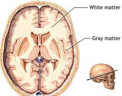
progressive multifocal leucoencephalopathy

Symptoms & Signs of Progressive Multifocal Leukoencephalopathy

Since the brain is the primary organ affected, the symptoms are reflective of brain dysfunction or even damage. Symptoms of progressive multifocal leukoencephalopathy include the following:

Sensory-Motor Changes

  1. Visual impairment or loss
  2. Tingling sensation or loss of sensation from the limbs
  3. Changes in hearing, smell and taste
  4. Non-reaction to a strong or painful sensory stimuli
  5. Speech Impairment or even aphasia (loss of speech)
  6. Inability to understand speech
  7. Unilateral or bilateral weakness
  8. Paralysis
  9. Loss of balance or clumsiness
  10. Seizures

Cognitive Changes

  1. Disorientation to time, place and people
  2. Progressive loss of memory
  3. Inability to focus or solve problems
  4. Inability to execute activities of daily living
  5. Progressive loss of consciousness
  6. Drowsiness

Symptoms of the disease occur in a quick and progressive manner. The symptoms stated above also resemble other more common neurologic conditions such as stroke, encephalitis or meningitis that is why it is crucial to immediately diagnose the condition and know the patient’s health history in order to delineate it from other acute conditions.

Diagnosis of Progressive Multifocal Leukoencephalopathy

DNA Sampling

To diagnose progressive Multifocal leukoencephalopathy, a DNA sample of the JC virus is taken from the cerebrospinal fluid. The cerebrospinal fluid (CSF) is a clear fluid that surrounds the brain and the spinal cord. In cases of infections, the CSF usually contains a number of these bacteria and viruses. To know if the neurologic condition is indeed caused by the JC virus, a sample of the CSF is taken where the DNA of the virus is extracted for verification. DNA sample can also be acquired through a biopsy of the brain tissue. In this procedure a small sample of the brain tissue is acquired and studied microscopically for the presence of the JC virus.

Magnetic Resonance Imaging

This is another way to diagnose progressive multifocal leukoencephalopathy (PML). An MRI gives an accurate image of the brain and the damages caused specifically by the PML. If MRI images show non-enhancing, multifocal lesions without mass effect, then the diagnosis for progressive multifocal leukoencephalopathy becomes evident.

Electroencephalogram or EEG

An EEG is also done to a patient suspected of having PML. This test is to evaluate the patient’s brain function and if there are any abnormalities present.

Blood Test and Urine Test

Taking blood samples and urine is done to test for the presence of the JC virus. High concentrations of the JC virus in the blood or urine may indicate an ongoing infection.

Treatment of Progressive Multifocal Leukoencephalopathy

Still, there is no known cure for progressive multifocal leukoencephalopathy. Some cases of PML resolve when the patient’s immune system improves, in cases of AIDS patients improvement of PML is seen with HAART or highly active retroviral therapy. Though there is no single cure for this condition, the following treatments are seen to improve and increase the survival rate of patients.

Antiviral agents

Interleukin-2 and cidofovir are antiviral drugs given to PML patients, these drugs act against the virus, destroying it and preventing it from reproducing.

Cytarabine

A chemotherapeutic drug used for cancer treatment but is seen to improve PML patient’s neurologic condition. According to results from clinical trials, this drug is seen to stabilize the neurologic condition of PML patients. in one case, it is seen to help a patient regain some cognitive function that was lost due to PML.

Mefloquine

An antimalarial drug is seen to eradicate the JC virus and prevent neurologic deterioration.

Prognosis of Progressive Multifocal Leukoencephalopathy

After diagnosis, a mortality rate of 30-50% occurs in patients with progressive multifocal eukoencephalopathy. But this is still dependent on the patient’s overall health, the treatment he/she receives and the underlying conditions that are present.

Patients who survive the disease can have neurologic disabilities, in which some cases are severe.

No responses yet

Focal Nodular Hyperplasia – Treatment, Symptoms, Causes

Aug 04 2015 Published by under Diseases & Conditions

What is Focal Nodular Hyperplasia of the Liver?

Focal Nodular Hyperplasia is a benign tumor growth in the liver. Focal nodular hyperplasia is the second most common benign growth in the liver after hemangioma. Most cases are asymptomatic and are only diagnosed accidentally from imaging studies, while one third of the cases are discovered due to the emergence of clinical symptoms.

This is a benign tumor growth, and has rare tendencies to become malignant, these benign lesions rarely causes bleeding and does not grow rapidly.

Characteristics

The tumor growth in focal nodular hyperplasia has the following characteristics:

  • Benign (no tendencies to be malignant)
  • Has hepatocytes or liver cells
  • Contains bile duct elements
  • Presence of fibrous tissue formation
  • Contains Kupffer cells
  • hepatocyte hyperplasia or the increase in the number of cells is evident

Clinical Presentation

Only a third of the cases of focal nodular hyperplasia shows symptoms. These cases may indicate an increased rate of cellular proliferation that is enough to cause signs and symptoms. Most cases are asymptomatic and are accidentally seen in X-Rays or CT scans.

When focal nodular hyperplasia is suspected or seen, it manifests the following clinical presentation:

  • Gross feature is a scar with a stellate center, when the tumor is dissected and examined; a star-like scar is seen at the center of the growth.
  • Large numbers of bland appearing hepatocytes proliferate the lobules of the liver And this is accompanied by the presence of bile compound and malformed blood vessels.
  • Telangiectasia is also seen, this is a condition where small blood vessels become dilated. This is often accompanied by cellular changes such as dysplasia.
  • Slower rate biliary secretion
  • Tumors may be present on the liver surface or pedunculated
  • Most lesions are smaller than 5cm most with an average diameter of 3 cm
  • In some cases the lesion replace a lobe of the liver, this is then called Lobar Focal nodular hyperplasia

How Common is Focal Nodular Hyperplasia?

This is a rare condition with the following prevalence:

  • 8% of liver tumors among adults is attributed to focal nodular hyperplasia. An approximate of 0.4-3% of the general population develops this condition. studies also reveal that the risk of focal nodular hyperplasia increases with age.
  • For children, 2% of liver tumors are attributed to this condition. an estimated 2.25 per million children are affected by focal nodular hyperplasia. Focal nodular hyperplasia ranks third in the pediatric liver tumor diseases.
  • Focal nodular hyperplasia is seen to occur in higher rates among females, but further studies are still done to explore this prevalence.

Symptoms and Signs of Focal Nodular Hyperplasia

This is usually an incidental finding, meaning the condition is only known accidentally when the patient underwent an imaging test for evaluation of another disease. A symptom are usually rare and includes the following:

  • Serum liver results that are abnormal
  • Fever
  • Abdominal enlargement (especially on the right side)
  • Palpable mass on the right upper abdomen
  • Hepatomegaly or enlargement of the liver
  • Weight loss

Causes and Pathogenesis

The exact cause of focal nodular hyperplasia is still unknown; however certain factors are seen to contribute to the formation of the benign lesions.

  • Abnormalities in the blood vessels – The exact mechanism to why a person develops focal nodular hyperplasia is unknown but an accepted theory to why the disease emerges is the presence of abnormalities in the blood vessels. In focal nodular hyperplasia, additional networks of blood vessels and arterial malformations are seen. These abnormalities can either feed the liver cells with too much nutrients or oxygen or could leave them deprived of both. When either over nourishment and undernourishment happens, the cells increase in number
  • Genetic abnormalities and inherited conditions – Focal nodular hyperplasia is also seen to occur in people with inherited conditions such as hemorrhagic telangiectasia. This is an inherited condition characterized by vascular malformations as well. This genetic predisposition causes vascular malformation in the liver and eventually leads to focal nodular hyperplasia.
  • Past childhood malignancy – If a child had been treated for a past malignancy, he/she is at a higher risk of developing focal nodular hyperplasia. This is observed in clinical studies where 138 patients who received treatment for malignancies developed focal nodular hyperplasia. The incidence rate is at 045% of the cases.

Diagnosis

  • Color Doppler – Focal nodular hyperplasia (FNH) can be diagnosed with the use of imaging studies such as a color Doppler. Upon diagnosis a focal nodular hyperplasia would appear like a “comet tail”. This is because FNHs are hypervascular in nature; upon Doppler studies scattered veins and arteries are seen throughout the liver, giving a comet like appearance.
  • Ultrasound – Ultrasound can also diagnose focal nodular hyperplasia since these tumors are isoechoic. But ultrasound studies can only detect tumors that are already pressing on to the adjacent structures or liver vessels. Ultrasound can only detect tumors with exerting mass effects.
  • Ultrasound with Enhanced Contrast – This imaging study can help diagnose FNH by showing through contrast the classic sign of focal nodular hyperplasia: a central lesion that is non-enhancing and resembles a stellar shape.
  • Low Index Contrast Enhanced Sonography – This diagnostic procedure enables the clinician to differentiate the condition from hepatic adenoma or other liver conditions. Focal nodular hyperplasia usually masks other liver conditions that need immediate or prompt treatment. Differential diagnosis to rule out focal nodular hyperplasia from any other forms of liver tumor formations is a priority intervention.
  • Computed Tomography or CT Scan – A CT scan is usually done to point out the location, size and density of the tumor growth. A CT scan of a focal nodular dysplasia usually shows deformity in the contour of the liver, or a liver with a star shaped scar in the center. CT scans are often employed to give an overview of the growth but it doesn’t tell much about the characteristics of the tumor itself.

Imaging Studies that Reveal Characteristics of Focal Nodular Hyperplasia

  • Nuclear Scintigraphy – This imaging study uses Sulphur colloid scan. 60% of focal nodular hyperplasia lesions would uptake this radiotracer, which is frequently seen in hepatic adenomas.
  • MRI with Superparamagnetic Iron Oxide (SPIO) – MRI imaging enhanced with SPIO is used to determine the characteristics of focal nodular hyperplasia lesions. Since the lesions contain Kupffer cells, the contrast medium (SPIO) is greatly absorbed by the cells, indicating that the lesion is FNH in nature rather than an adenoma.

Treatment of Focal Nodular Hyperplasia

Resection of Focal Nodular Hyperplasic Liver – Surgery remains to be the primary mode of treatment. This is true for patients found to have the condition upon incidental finding or for those showing pronounced symptoms. Surgery involves the removal of the affected area (the tumor and some liver tissue or the entire lobe of the liver). After surgery follow up checkups are indicated to evaluate emergence of new tumors and the progression of their growth.

Indications for Surgery

This condition is rare and for most treatment nor surgery is not indicated. However on certain instances, a liver resection may be indicated:

  • Lesions that are multiple
  • Large lesions, probably greater than 5cm
  • Liver enlargement or displacement due to the tumor formation
  • Symptom producing tumors
  • IF the behaves abnormally or increases size rapidly
  • If there are signs of intra-lesional hemorrhage

Surgery involves liver resection. Liver resection is the removal of a part of the liver that is diseased or affected. In focal nodular hyperplasia, the part of the liver where there is tumor growth is removed. The healthy part of the liver is left unharmed. A person can still continue normal functioning despite the removal of one part of the liver.

Moderation in alcohol, medications and fat intake must be observed to prevent straining the liver. Of the right part of the liver is affected, the gallbladder may also be removed. Still if the removal of the gallbladder does not warrant a threat in one’s life. But dietary modifications and changes in lifestyle would be necessary.

No responses yet

© 2017 HealthFoxx.com. All Rights Reserved. Privacy Policy
This website is for informational purposes only and Is not a substitute for medical advice, diagnosis or treatment.