Archive for October, 2013

Erythema Annulare Centrifugum

Oct 27 2013 Published by under Diseases & Conditions

Whay is Erythema Annulare Centrifugum?

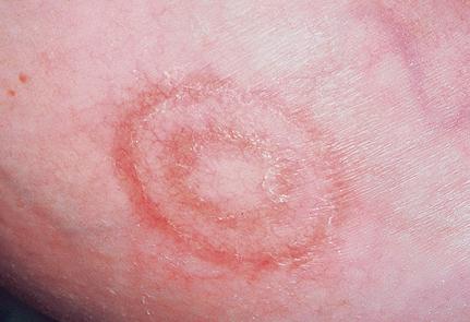
Erythema annulare centrifugum is a term used to describe skin lesions. It was first described by Darier in 1961 and is characterized as nonpruritic, scaling or nonscaling, red in color, ring form or can be curved. Lesions spread from the center and may may last from a few days to a few months. Other names for Erythema annulare centrifigum are”deep gyrate erythema,” “Erythema perstans,” “Palpable migrating erythema”, and “superficial gyrate erythema.”

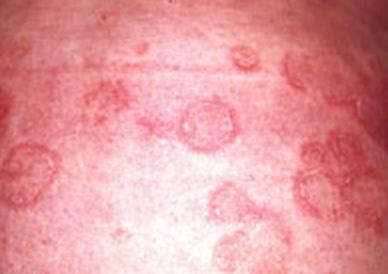
Lesions usually begin as a small pink to red spot, ultimately becoming bigger at a rate of about two to five millimeters per day and extend up to six to eight centimeters. Lesions form in a ring shape while its central area flattens and clears. Sometimes they grow into irregular shape. Eruptions start to appear on the thighs and legs but may also appear on the face, chest and upper extremeties. EAC is usually without symptoms but sometimes mild itching is experience by the patient. It is considered as rare disease, affecting one in 100,000 per year.

Erythema Annulare Centrifugum 1

Causes

According to studies, Erythema annulare centrifugum has been linked with different etiologic factors. Erythema annulare centrifugum is also associated with underlying diseases and conditions. These include:

Bacterial, fungal, filarial and viral infections

Example of these are tuberculosis, sinusitis, lyme disease, candidiasis or tinea.

Medications

Hypersensitive reactions to a variety of drugs such as antimalarials (chloroquine and hydroxychloroquine), aldactone, oestrogen, penicillin and amitriptyline.

Cancer

Carcinoma, blood dyscrasias

Food

Ingestion of blue cheese or tomatoes

  1. Ascaris Infestation
  2. Arthropod bites
  3. Pregnancy
  4. Hormones
  5. Idiopathic or an unknown cause
  6. Other diseases such as recurrent or chronic appendicitis, cholestatic liver disease (blocked bile system), and Graves disease (overactive thyroid gland) and autoimmune disorders like Lupus.

Histopathology

In classic or deep cutting in the middle and lower dermis, an intense, superficial and deep lymphocytic or lymphohistiocytic perivascular infiltrate in a coat-sleeve fashion. While the superficial type, the presence of more nonspecific perivascular lymphohistiocytic infiltrate the superficial dermal vessels and edema of the papillary dermis is noted.

A study made by medical practitioners revealed the substantial differences between cases with a wholly superficial type and cases with a superficial and deep infiltrate. Clinically, a collarette of scales was seen only in the superficial type. Histopathologically, some findings were much more common in the superficial type (eg, spongiosis, parakeratosis, crusts, edema of the papillary dermis, epidermal hyperplasia) and in the deep type (eg, sleeve-like arrangement of the infiltrate, melanophages, subtle vacuolar changes at the dermo-epidermal junction, individual necrotic keratinocytes). Whereas cases of the superficial type could be distinguished from differential diagnoses by a variety of clinical and histopathologic findings, most cases of the deep type showed subtle signs of lupus erythematosus. Neither type was associated consistently with any other systemic disease. Because the superficial and the deep type of erythema annulare centrifugum seem to be unrelated to one another, they should not be referred to by the same name.

Treatment

EAC is usually self-limiting, ie. it clears up by itself. Determining the appropriate treatment and ruling out underlying disorders is the primary therapy.

Topical steroids

Used to relieve swelling, redness and itchiness. However, topical steroids are not proven to be effective to prevent the occurrence of new lesions.

Injection steroid therapy

Effective in reducing inflammation, but recurrence of lesions happen after stopping the therapy.

Topical calcipotriol

A topical vitamin D derivative has been known to be beneficial.

ICD-9 Code

The International Statistical Classification of Diseases and Related Health Problems or ICD is a system published by World Health Organization (WHO) that provide codes in which every health condition is assigned to unique category. ICD is used worldwide to classify diseases and different kinds of signs and symptoms, abnormal findings, physical assessment, social circumstances, and external causes of injury or disease. ICD is also use to determine the morbidity and mortality percentage.

Erythema Annulare Centrifugum

L53.1 is a billable ICD-10-CM code that can be used to specify a diagnosis.
ICD-10-CM will officially replace ICD-9-CM on October 1, 2013, therefore, L53.1 and all ICD-10-CM diagnosis codes should only be used for training or planning purposes until then.

Is Erythema Annulare Centrifigum Contagious?

At present, EAC is not known to be contagious. No studies have been proven that EAC is contagious.

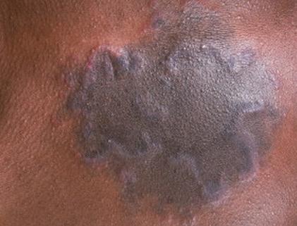
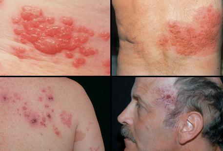
Erythema Annulare Centrifugum pictures

photos, pics, images of Erythema Annulare Centrifugum

Erythema Annulare Centrifugum picture

Erythema-Annulare-Centrifugum-image

Erythema-Annulare-Centrifugum-photo

No responses yet

Foraminal Stenosis – Types, Causes, Symptoms, Diagnosis, Treatment

Oct 23 2013 Published by under Diseases & Conditions

Foraminal stenosis is the narrowing of one or more vertebral foramen. Vertebral foramen refers to the opening between the anterior segment and the posterior segment of the vertebral column, allowing nerve roots to pass through it. Vertebral foramen is located on the left and right side of the spine. Vertebral foramen is placed from the cervical spine up to the lumbar spine.

Definition of Foraminal Stenosis

Foraminal stenosis involves the reduction in size of the vertebral foramen that allows the nerve roots to pass through it. Once the space has decreased because of stenosis, the nerve roots become compressed.

Foraminal Stenosis Symptoms

Image Source: http://www.backcare101.com

Types of Foraminal Stenosis

Foraminal Stenosis has varied types depending on the location, severity and area of narrowing.

Types according to the area of narrowing:

  • Bilateral Foraminal stenosis

Bilateral foraminal stenosis involves the narrowing of the foramina on both sides of the spine. Foramen is located on both sides of the spine and the narrowing of both sides causes bilateral foraminal stenosis.

  • Unilateral Foraminal stensois

Unilateral stenosis only involves the stenosis on the left of the spine

  • Far Lateral stenosis

This involves narrowing where the nerve root has exited the foramen.

Types according to Location

Cervical

Cervical stenosis is the most common form of foraminal stenosis. The affectation of the nerve roots in the area commonly affect the neck, shoulders, head, arms, hands and upper back.

Thoracic
Thoracic stenosis is an uncommon form because this area is usually protected from degenerative diseases. When this area is affected, common locations include the ribs, arms, shoulders and internal organs.

Lumbar
Lumbar stenosis is also a common form of foraminal stenosis affecting the lower extremities, buttocks, and lower back.

Multilevel
This involves the narrowing of the foramen at different levels in the spine. This is a more severe type because it can affect one or more areas in the body.

Types according to severity

Mild
Mild foraminal stenosis primarily results from natural aging. Some cases are asymptomatic and may not be detected.

Moderate
Moderate foraminal stenosis or neural foraminal stenosis is the gradual narrowing of the passage ways of nerve roots. This also involves more evident symptoms such as pain and alterations in senses in the extremities or any area of affectation. Neural foraminal stenosis involves the significant compression of nerves as compared with mild stenosis. This is usually caused by disc degeneration.

Severe
Severe foraminal stenosis is a degenerative condition that causes severe pain in the back or neck. This results from wear and tear that the spine experiences and is usually exacerbated by injury.

Causes of Foraminal Stenosis

Several factors cause clogging or narrowing of the vertebral foramen. These include:

Disc degeneration

This is the most common cause of foraminal stenosis as a result of natural aging. Disc degeneration usually develops after disc herniation or the bulging of the disc outside its cavity.

Rheumatoid arthritis

The occurrence of this degenerative condition as a result of rheumatoid arthritis causes the foramen to narrow. This is caused by the thickening of the joint tissue, calcification of the spine, bone spurs, and inflammation in the area.

Osteoarthritis

This is one of the most common causes of cervical foraminal stenosis. This results from the degeneration of the spine as a result of its weight-bearing function. Osteoarthritis involves damage to the cartilage on the spine’s joints that may cause bone spurs that may impede the passage of nerve roots.

Congenital spinal stenosis

Narrowing of the vertebral column that appears during the fetal development may also cause foraminal stenosis.

Scoliosis

Scoliosis is a condition wherein there is lateral curvature of the spine. This may affect one side of the vertebrae and lead to unilateral foraminal stenosis.

Aging

Increasing age also plays a role in the development of foraminal stenosis. As people age, the spinal integrity becomes affected, leading to narrowing of the foramen.

Repetitive Activity

Repetitive tension on the spine as a result of high impact activities can also lead to bone changes such as calcifications and bone growths that obstruct the nerve passage.

Symptoms of Foraminal Stenosis

Symptoms of foraminal stenosis occur because of compression of the nerve roots. Nerve roots innervate specific areas of the body and these areas may be affected. Some of these include:

  • Pain on the area affected such as back pain, leg pain or shoulder pain
  • Weakness
  • Numbness
  • Diminished reflexes
  • Paresthesia or the feeling of tingling and pins and needles in the affected area
  • Loss of function as a result of an untreated condition

Pain can also be characterized as radicular, which means that there is the presence of pain along the dermatomes and the affected nerve root areas. Pain can be characterized as sharp and aching.

Diagnosis of Foraminal Stenosis

Diagnosis of foraminal stenosis is done using the following procedures:

Physical examination

Physicians often perform physical examination, focusing on the assessment of sensations such as touch, pain and temperature. The reflexes are also checked and the examiner will also assess for muscle strength.

Imaging studies

X-rays, CT scans, and MRI scans be done to ascertain the diagnosis. These tests provide a view of the spine and the extent of the compression and narrowing. These also determine the extent of treatments needed, from conservative anti-inflammatory medications to surgery.

Treatment of Foraminal Stenosis

The treatment for foraminal stenosis depends on the extent of symptoms and the narrowing. Several methods can be employed, including:

Foraminotomy

This is a surgical procedure that employs the creation of an opening on the blocked foramen, used for more severe cases of narrowing.

Selective Nerve Root Block

This involves temporary pain relief by injecting anesthetics in the spine in the affected area to relieve pain. This treatment is also called selective transforaminal epidural injection.

Physical Therapy

To improve the mobility of the patient and reduce symptoms, physical therapy may be employed as a conservative management for this condition.

Rest

Rest is an important treatment method to prevent further damage and nerve compression. In cases of severe stenosis that cause severe pain, immobilization may sometimes be required.

Anti-inflammatory Drugs

Medications that relieve inflammation are also given to reduce the inflammatory process in the area and reduce nerve compression. Anti-inflammatory medications can be in the form of non-steroidal anti-inflammatory drugs or corticosteroids.

No responses yet

Gestational Trophoblastic Disease – Symptoms, Diagnosis, Treatment

Oct 18 2013 Published by under Diseases & Conditions

What is Gestational Trophoblastic Disease?

Gestational Trophoblastic Disease is a rare tumor that occurs during pregnancy. This kind of tumor starts with the trophoblast area, a cell that provides nutrients to the embryo as well as contributing in the formation of the placenta. Normally, when this disease is present, child development is impossible.

Types

There are four main types of Gestational Trophoblastic Disease which are divided into two categories:

A) The Benign Tumor

Molar Pregnancy

It is the most common type of Gestational Trophoblastic Disease where there is an abnormal mass growth inside the uterus. It is a result of having an over-production of tissue in the placenta where the newborn is usually present. This molar pregnancy is divided in to two types:

  1. Partial Molar Pregnancy develops when two sperms fertilize the egg. It means that there is abnormal placenta and some development of  the fetus.
  2. Complete Molar Pregnancy develops when one or two sperms fertilize the empty egg cell. There is an abnormal placenta but no fetus is formed.

Signs and Symptoms:

  1. There is an abnormal growth of the uterus and some women might think they are pregnant
  2. Nausea and vomiting
  3. During first three months of pregnancy there will be some cases of vaginal bleeding
  4. Feeling of restlessness, increased heart rate, heat intolerance and unexplained weight loss.
  5. High blood pressure and swelling of the extremities.

B) The Malignant Tumors

B1. Invasive mole

It is a tumor where there is an invasion in the uterus that also spreads and metastasizse in other parts of the body.

Signs and Symptoms:

  1. Abdominal swelling
  2. Excessive vomiting
  3. Fatigue often caused by anemia due to severe vaginal bleeding
  4. Abdominal pain
  5. Pelvic pain and discharge from the vagina
  6. In rare cases, the patient will feel shortness of breathe as well as coughing up blood due to the invasion of cancer cells in the lungs.

B2. Choriocarcinoma

It is a type of cancer that grows rapidly in woman’s uterus. With this cancer, there is a big possibility that even after removing the mole, it can continue to spread and grow again into cancer.

Signs and Symptoms:

  1. Vaginal bleeding
  2. Uneven swelling of the uterus
  3. Abdominal and pelvic pain

B3. Placental-Site Trophoblastic Tumor

It is a rare form of Gestational Trophoblastic Disease that develops in the area where the placenta attaches to the uterus. It is a slow-growing tumor and doesn’t easily invade other parts of the body. It is also a tumor that is easily curable.

Signs and Symptoms:

  1. Normally, it is asymptomatic in nature
  2. Abnormal vaginal bleeding
  3. Enlarged uterus
  4. Unusual pain in the abdominal area

Causes

  1. The exact cause is unknown
  2. It is normally associated with (1) age : women above 40 years old and below 20 years old. (2) Previous molar pregnancy, (3) miscarriage, (4) blood type: RH incompatibility, (5) women who take pills, (6) diet, (7) family history.

Diagnosis

  1. Family and medical history including age and history of previous abortions or miscarriages.
  2. Physical examination if there is any medical condition that might affect diagnosis of the disease. This includes vaginal bleeding, abdominal and pelvic pain.
  3. Urine and blood samples to determine if there are any abnormal hormons present that relate to the formation of the disease.
  4. A biopsy is a medical test done by professionals where they will take a tissue sample for examination. The sample is checked through a microscope and helps to determine any diseases.
  5. Computed Tomography Scan (CT scan) is an X-ray that can provide sharper and more detailed images of the lungs. It combines a series of X-ray views with different angles that produce cross-sectional images of bones and tissues.
  6. MRI or Magnetic Resonance Imaging is also like the CT scan and helps detect any formation or abnormal growth in the body, but it is clearer because it uses a dye to see images.
  7. Ultrasound that can also help in viewing internal organs and determine any abnormal tissues.

Treatment

  1. Suction Curettage – a procedure that suctions the contents of the uterus through the cervix. Normally, it is used for abortion purposes, but it also aids in regulation of menstruation and is  used to terminate any molar pregnancy
  2. Hysterectomy – a surgical procedure where surgeons remove the uterus. Usually, it is recommended to women who don’t want to have future pregnancies. It is recommended if there is no other available treatment available.
  3. Chemotherapy – a procedure done usually after surgery to eliminate possible cancer cells that are not removed in surgery. There are two types of chemotherapy done to patients with Appendix Cancer. These are:
  4. Local Chemotherapy – this type of chemotherapy focuses on one section of the body and is the common type of chemotherapy used in this disease. Medical professionals normally remove the accumulated fluid inside the patient’s stomach before doing this procedure in order for the chemotherapy to be effective. A tube is inserted after cytoreduction surgery to deliver the chemotherapy procedure. Normally, it is done only once.
  5. Systemic Chemotherapy – a type of chemotherapy procedure that is delivered to the bloodstream to reach cancer cells that have scattered in the body. This type of chemotherapy needs many cycles and the effect of this therapy depends on the person and the dose given. The patient often experiences fatigue, nausea, vomiting, and loss of appetite but these symptoms subside eventually after the treatment.
  6. Radiation Therapy – a therapy used in patients using high-energy X-rays to kill cancer cells. This type of therapy is not a common treatment for patients with appendix cancer. This type of therapy is delivered in many cycles. It is used to kill cancer cells without affecting the tissues that surround the cancer cells; it can also used as a treatment to reduce the size of tumors and reduce the amount pain.

No responses yet

PostHerpetic Neuralgia – Treatment, Symptoms, Causes, Prevention

Oct 14 2013 Published by under Diseases & Conditions

What is Post-Herpetic Neuralgia?

PostHerpetic Neuralgia is a complication of Herpes-Zoster which is defined as a painful condition that normally showsup  when the Herpes-Zoster is already in the healing process. Herpes-Zoster is a viral disease, causing painful rashes and accompanied by blisters. Normally, it affects elderly people, aged 50 years old and above.

Shingles Pictures slideshow

Source – medicinenet.com

PostHerpetic Neuralgia Signs and Symptoms

  1. Pain for three months or more
  2. Stabbing or burning pain
  3. Rashes on the chest, forehead, stomach, or any parts of the body
  4. Formation of blisters that eventually will heal but pain lingers
  5. People with PostHerpetic Neuralgia are sensitive to touch because of the sensitivity of the nerves
  6. People who have this disease are also sensitive to temperature changes.
  7. Feeling of numbness if the nerves involved control the movement of an individual
  8. Itchiness and headaches

Causes

PostHerpetic Neuralgia is caused by Varicella Virus which is also is the virus present in the chicken pox, but the difference is that chicken pox is normally seen in children while PostHerpetic Neuralgia is seen in older adults. When a person has had chicken pox, some of the virus remains in the nerve cells of the body. Years later when the person’s immune system decreases, it can possibly reactivate the virus and lead to the formation of Herpes-Zoster. This disease usually occurs when the nerve fibers in our body become damaged during an occurrence of Herpes-Zoster. This disease is due to changes in the nervous system caused by complications of Herpes-Zoster. Since this disease is caused by the development of the Variccela Virus reactivated during the healing stage of Herpes-Zoster, the damaged fibers have difficulty sending messages from the skin to the brain that causes the “pain” message to be disorientated and overstated, resulting in burning, piercing pain that the person will experience for months.

Pathophysiology

  • Stage 1 of Varicella Infection: chicken pox during childhood
  • Stage 2 of Varicella Infection: latency period or where the Varicella Virus is in state of inactivity
  • Stage 3 of Varicella Infection: reactivation of Varicella Virus: Herpes-Zoster – in older adults as their immune system decreases
  • Stage 4 of Varicella Infection: PostHerpetic Neuralgia – during the healing stage of Herpes-Zoster Infection

PostHerpetic Neuralgia Treatment

Treatment for this disease will depend on the pain the person experiences and patient’s physical and neurologic health. These treatments are:

Non-Pharmacological Treatment

  1. Use of loose-fitting clothing to reduce the irritation caused by the disease.
  2. Pain may eased by cooling the affected area like ice cubes wrapped in a cloth or plastic or by giving the patient a cool bath.
  3. Relaxation Techniques are used to distract the mind from the pain. It helps the person to relax, to reduce levels of anxiety and stress as a result of the pain.
  4. Heat therapy as a rehabilitation purpose that can decrease pain, reducing the inflammation and increase blood flow that will provide better healing.
  5. Nerve stimulation is a type of treatment using a small amount of electricity to stimulate the nerves for healing purposes. It is normally recommended by medical professionals to treat pain.
  6. Spinal Cord Stimulator is a device used on patients to send electrical signals in the spinal cord to help to control the pain.

Pharmacologic Treatment

  1. Analgesics is known to relieve pain. Some analgesic used are aspirin, lidocaine patches and paracetamol.
  2. Antidepressants is helpful to cure both depression and pain. This type of medication is given to patients in a low-dosage compared to patients with depression. Most of the anti-depressant drugs don’t eliminate pain, but they make the pain tolerable.
  3. Anticonvulsant medication is used for patients to treat muscle spasms and help in pain modulation. .

Prevention

The best method to prevent the disease is to have early treatment when Herpes-Zoster appears. Contact a medical professional once there are signs of Herpes-Zoster to reduce your chance of having the disease. The best way to prevent PostHerpetic Neuralgia is vaccination against chicken pox as well as Herpes-Zoster.

  1. Chicken pox vaccine is given to the children between the age of one year and one year four months, but it can also be given to adults who have never experienced chicken pox. This type of vaccine doesn’t means that it can provide 100% protection against the disease, but it can lessen the possible risk of complication and it can reduce the severity of the disease.
  2. Herpes-Zoster vaccine can help older people who have already experienced chicken pox. Like the chicken pox vaccine, it doesn’t guarantee 100% protection against the virus. It can lessen the possible risk of complication and help in reducing the severity of the disease. The vaccine is used only for protection, not for treating people who are already infected. People aged 60 and above are advised to have this vaccine to prevent complications due to a decreased immune system that might reactivate the Varicella Virus and eventually lead to Post-Herpatic Neuralgia

No responses yet

Anion Gap Metabolic Acidosis – Low & High Causes, Symptoms

Oct 08 2013 Published by under Diseases & Conditions

What is Anion Gap Metabolic Acidosis?

The Anion gap is the difference in the cations and anions in the body. Cations are positively-charged ions while anions are negatively-charged ions. The serum anions and cations are usually used, but urine measurements can also be used. The degree of the gap in the serum electrolytes is calculated to determine reasons for metabolic acidosis. When the gap is higher than normal, high anion gap metabolic acidosis is revealed, whereas if the gap is low, low anion gap metabolic acidosis is concluded.

The anion gap refers to all the unmeasured anions in plasma. Commonly measured cations include sodium, potassium, calcium and magnesium. Cations that are normally unmeasured include some pathological proteins. Commonly measured anions include chloride, phosphate, and bicarbonate and commonly unmeasured ones include sulphates, lactate and acetoacetate. The unmeasured anions account for about 10% of anions in the plasma. The anion gap represents these unmeasured anions which are not part of the usual laboratory tests.

When metabolic acidosis occurs, the hydrogen ions (cations) reacts with bicarbonate anions, resulting in decreased concentration of the measured anion and increased concentration in the unmeasured anions, leading to a gap.

Anion gap is used for the following reasons:

  1. To signal the presence of metabolic acidosis and confirm other findings
  2. To differentiate the causes of metabolic acidosis
  3. To assess the extent of metabolic acidosis and help in treatment choice.

Anion Gap Levels

Anion gap is subdivided into levels depending on the symptoms and cause.

Low Anion Gap

A low anion gap includes a measurement of less than three mEq/L. It is an infrequent form of metabolic acidosis and accounts to only one to three percent of all cases.

Causes

A low anion gap is usually caused by hypoalbuminemia, a decrease in albumin in the blood. Albumin is an anion which when decreased, it allows other anions such as bicarbonate and chloride to be retained. This causes the gap to decrease.

Common conditions that lead to a low anion gap are hemorrhage, liver cirrhosis, nephritic syndrome, and intestinal obstruction. Multiple myeloma also may cause a low anion gap because of an increase in plasma immunoglobulins that leads to hypoalbuminemia.

Symptoms

Symptoms include:

  1. Hypoalbuminemia
  2. Hypercalcemia/ Hypermagnesemia
  3. Muscle weakness
  4. Edema
  5. Hypotension
  6. Lethargy
  7. Dysrhthmias or irregularities in the heart rate

Normal Anion Gap

In normal anion gap, the computed value is three mEq/L to 11 mEq/L.

Causes

In a normal anion gap, the drop in bicarbonate ions (HCO3¯) is compensated by an increase in chloride (Cl¯) which makes the gap remain normal despite of metabolic acidosis. It is also known as hyperchloremic acidosis

Common conditions that lead to a normal anion gap include:

  1. Diarrhea
  2. Alcoholic intoxication
  3. Addison’s disease
  4. Renal dysfunction (renal tubular acidosis)
  5. Uretero-enterostomy
  6. Saline administration
  7. Hyperparatrhyroidism
  8. Intake of carbonic anhydrase inhibitors (Acetazolamide) and ammonium chloride

Symptoms

Symptoms include:

  1. Increased serum chloride levels
  2. Fatigue
  3. Postural hypotension
  4. Emotional disturbances
  5. Increased thirst
  6. Anorexia
  7. Polyuria
  8. Restlessness
  9. Weakness

High Anion Gap

A high anion gap is characterized by a gap of more than 10 to 11 mEq/L.

Causes

In a high anion gap, the presence of acidosis causes the bicarbonate ions to decrease. Common conditions that lead to a high anion gap include the following:

  1. Lactic acidosis
  2. Ketoacidosis as seen in diabetes and alcoholism
  3. Dehydration
  4. Salicylate toxicity that results in metabolic block
  5. Medication overuse of isoniazid and Toluene
  6. Ingestion of toxins such as ethylene glycol, paraldehyde, methanol, propyl alcohol, Phenformin, cyanide, iron
  7. Renal failure because of a decrease in the ability of the kidneys to produce bicarbonates to buffer the acidosis.
  8. Uremia
  9. Rhabdomyolysis

Symptoms

Symptoms include:

  1. Low bicarbonate levels (less than 22 mEq/L)
  2. Fruity or acetone breath
  3. Warm, dry skin
  4. Abdominal pain
  5. Polyuria or increased urination
  6. Tachycardia
  7. Nausea and vomiting
  8. Diarrhea
  9. Hyperventilation as a compensatory mechanism
  10. Tinnitus or ringing of the ears as a result of salicylate toxicity

Diagnosis

Anion gaps are measured by computing the difference of the measured cations and anions in the body. Measurements are taken through blood tests. Anion gap is expressed in milliequivalents per liter (mEq/L) or millimoles per liter (mmol/L). By practice, only sodium, potassium, chloride and bicarbonate ions are used when calculating the anion gap.

Anion Gap Calculator- How to Calculate Anion Gap

The anion gap is calculated using two accepted formulas.

With Potassium:

Anion gap= ([Na+] + [K+]) ? ([Cl?] + [HCO3?])

Without Potassium:

Anion gap== [Na+] ? ([Cl?] + [HCO3?])

The calculation without potassium is usually used in daily practice. Since serum potassium is only low, the removal of it in the equation normally does not cause any significant result.

Blood Test for Anion Gap

In order to calculate the anion gap, blood testing for serum electrolytes (Na, K, Cl) and arterial blood gases (ABG) determination is used to check bicarbonate levels (HCO3). Patients should be instructed on monitoring electrolytes at regular intervals to assess extent of illness and efficacy of treatment. Specific electrolytes should be requested by the physician to be checked in order to keep costs down.

Treatment

Treatment of anion gap depends on the level and the underlying cause. Treatment goals are directed at managing the metabolic acidosis and include the following:

Administration of sodium bicarbonate therapy

To buffer the acidosis, bicarbonates need to be administered. The intravenous site needs to be checked because bicarbonates causes tissue irritation.

Electrolyte replacement of those with reduced levels

Serum sodium and potassium needs to be replaced in cases of hyponatremia or hypokalemia.

Administration of diuretics when dehydration is not present

This decreases serum calcium and magnesium levels. Diuretic used in cases of hypovolemis may further aggravate the condition.

Phosphate therapy

Phosphate enhances the elimination of excess calcium, magnesium, and chloride ions.

Albumin replacement therapy for hypoalbuminemia

Serum albumin is replaced by administering intravenous infusions.

Continuous monitoring of arterial blood gases, electrolytes, creatinine and BUN

Monitoring is done to determine treatment efficacy.

Prevention

Prevention of anion gap metabolic acidosis also depends on the cause. Prevention should be catered towards managing the disease to prevent metabolic acidosis. The following outlines the most common prevention for certain causes of metabolic acidosis.

Diabetic Ketoacidosis

  1. Take insulin as prescribed
  2. Monitor blood sugar frequently
  3. Prevent infection to avoid increase in insulin requirements
  4. Monitor hyperglycemia symptoms such as anorexia, nausea, vomiting and diarrhea for immediate management

Toxicity

  1. Take salicylates, anhydrase inhibitor diuretics, iron and isoniazid as prescribed
  2. Avoid exposure to cyanide and other toxins
  3. Drink alcohol in moderation

Lactic acidosis

  1. Avoid fatty foods to lower the risk of atherosclerosis that leads to anaerobic metabolism when it obstructs heart and brain circulation
  2. Watch out for hyperglycemia
  3. Exercise regularly to promote circulation

High Anion Gap Complications

Complications of anion gap usually involve the end results of metabolic acidosis such as:

  1. Coma
  2. Cerebral edema
  3. Respiratory collapse
  4. Cardiac dysrhythmias
  5. Hypovolemic shock or circulatory collapse

High Anion Gap Remedies

Metabolic acidosis is a highly complicated condition that requires full medical management. Remedies are not sufficient to treat the disease; however, it may improve some of the signs and symptoms. They include:

  1. Consume egg whites with every meal to replace albumin in case of albuminemia. Egg whites are a rich source of albumin.
  2. Increase intake of potassium rich foods such as bananas, apples and apricot to restore potassium levels.
  3. Consume sodium rich foods in the form of root crops.
  4. Encourage pursed lip breathing to allow for maximum exhalation of carbon dioxide, reducing carbonic acid levels.
  5. Increase fluid intake to manage hypovolemia.

Despite these remedies, patients with metabolic acidosis need medical management to prevent life-threatening complications. See for Anion gap calculation

No responses yet

Malabsorption Syndrome – Causes, Symptoms, Treatment, Prognosis

Oct 05 2013 Published by under Diseases & Conditions

Malabsorption syndrome is a collection of signs and symptoms that alters the ability of intestines to absorb nutrients and minerals especially fat-soluble vitamins A, D, E, K and macronutrients. The intestine cannot absorb properly because of the underlying conditions associated with this.
Malabsorption has many causes including bowel resection, vascular impairment, gallbladder disease and lymphatic disease. This condition can be acquired or congenital that really affects the transportation of nutrients in the body.

Condition Leading to Malabsorption Syndrome

1.) Celiac Disease– also known as gluten-sensitive enteropathy. It is a disease of the small intestine mark by the atrophy of villi and microvilli. It develops to an immune-mediated inflammatory response to gluten. Gluten is a protein commonly found in barley, rye, oats and wheat. The cause of this disease is unknown.
2.) Short bowel syndrome– happens when large portion of the small intestine is remove. It minimizes the area that absorbs nutrients.
3.) Lactose intolerance- is a condition wherein a person cannot tolerate in taking those products that contains lactose especially in milk.
4.) Whipples Disease– this condition is mostly affecting men in middle ages. It is rare condition that cause by bacterial infection.
5.) Topical sprue- a rare condition that is caused by food toxins.
6.) Short bowel syndrome- this condition lessens the surface area of the bowel that is responsible for nutrient absorption.
7.) Pancreatitis- this is a condition where the pancreas is infected and cannot absorb nutrients.
8.) Cystic fibrosis- the second widest condition causing malabsorption syndrome. This an autosomal recessive disorder affecting the exocrine glands.

Factors Affecting Malabsorption Syndrome

Several risk factors are being studied to know the morbidity of the syndrome.

  • Age (newborn and infants are most affected)
  • Race (common in United States, Eskimos in Canada, Greenland, Asia and Africa)
  • Sex (females highly acquire this condition)
  • Family history
  • Usage of specific drugs
  • Surgery
  • Excessive alcohol intake

Malabsorption Syndrome overview

Signs and Symptoms

  • Fatigue
  • Steatorrhea, Diarrhea, Abdominal distention, Foul-smelling stools, Abdominal bloating
  • Anemia, Edema
  • Musclecramps, Muscle Wasting
  • Skin irritation
  • Abnormal Heart rate
  • Low-grade fever
  • Pain in the joints
  • Weight loss
  • In some cases, radiation therapy is considered one of the reasons in this condition. Also antibiotics can affect a normal bacterium that resides in the intestine.

Laboratory and Diagnostic Testing

Some tests are required to perform in order to know and diagnose the condition. These test were listed here as follows:

  • Blood testing such as:

-serum sodium, potassium and chloride
-serum calcium and serum cholesterol
-serum protein and albumin test
-vitamin A indicator test (conforming beta-carotene)

  • Schilling test (skin testing)
  • D-xylose test
  • Fluids and fluid retention monitoring test
  • Electrolytes balance monitoring
  • Magnetic Resonance Imaging (MRI)
  • CT scan is also done
  • Barium enema
  • Stool collection monitoring for 72 hours
  • Biopsy of the intestine
  • X-ray of gastrointestinal tract
  • Shortness of breath

Treatment Modalities

Treatment also depends on the underlying cause. Different approach is done to evaluate the effectiveness of the treatment. These are the following:

  • Careful monitoring is done to prevent further medical illness
  • Essential vital signs is established
  • Assess for any signs of nutrients and mineral depletion
  • Hospitalization is needed if nutrient imbalance occurs.
  • Symptomatic approach is perform to alleviate discomforts
  • Oxygen is administered for shortness of breath
  • Dietary modifications
  • Supplemental vitamins and minerals are needed
  • Monitor frequently the growth of the patient
  • Monitor characteristics of stool and frequency of it
  • Supportive treatment of family is recommended
  • Daily oral intake of iron and folic acid is instructed to the patient
  • Anti-spasmodics to lessesn the muscle cramping
  • If the patient have cystic fibrosis and pancreatitis they are given pancreatic supplement
  • Dietary modification especially those who have lactose intolerance are instructed
  • Careful monitoring to ensure the stable condition of the patient
  • The expected outcome of this treatment varies on the prognosis of the person. Following proper treatment regimen is also recommended for the success of the treatment. Positive outlook in life can be a big help in controlling and minimizing the disease.

Disease Prognosis and Mortality

The frequency of this syndrome depends upon the underlying diseases. About 1% of the population can manifest this syndrome. This is not only by genetic means but this can also be acquired most especially by children of young age. Best to describe is protein that comes from cow and soy milk. It contains this macronutrient that a young kid cannot absorb properly. This on and off occurrence can lead to enteritis. This is another form of inflammation in the colon. A public demonstration of this condition differs in line with the specific disease condition. Depending on the particular disease, the mortality would be determined. If it is acquired congenitally, it will occur about 24 to 25 out of 100,000 live births with the highest mortality rate. The underlying should be determined first before it’s too late for the affected person to survive.

References:

  • http://emedicine.medscape.com/article/931041-overview#a0199
  • http://www.faqs.org/health/topics/3/Malabsorption-syndrome.html#xzz2f1Jby0VZ
  • http://medical-dictionary.thefreedictionary.com/Malabsorption+Syndrome
  • http://www.nlm.nih.gov/medlineplus/malabsorptionsyndromes.html

No responses yet

Reactive Airway Disease in Children, Adults – Symptoms and Treatment

Oct 02 2013 Published by under Diseases & Conditions

What is Reactive Airway Disease?

Reactive Airway Disease is a condition involving the antigen-antibody reaction in the lung and the activation of the inflammatory cascade that results in airway inflammation. Reactive airway disease should not be confused with asthma, as the disease is broader than asthma in children and adults. Patients may experience wheezing without asthma. However, the manifestations and management of reactive airway disease and asthma is synonymous.

Reactive airways disease affects up to one-third of the population in children. Reactive airway disease accounts for up to 200,000 hospitalizations and 13 million healthcare visits in the United States. Reactive airway disease is common in Hispanic and black children. In fact, hospitalizations from reactive airway disease is four times higher in African Americans that in whites. Reactive airway disease also affects both children and adults with the peak of affectation in children during the ages six to eleven.

The mechanism of the disease involves narrowing of the airways as a result of bronchoconstriction and airway inflammation with resulting production of lung secretions. The changes in the airways results from the activation of an inflammatory response and immunity because of an antigen inhaled into the lungs.

Reactive Airway Disease

Symptoms of Reactive Airway Disease

Reactive airway disease symptoms result from the inflammatory cascade that happens in the lungs. Asthma may also be a sign of reactive airway disease, making the two interchangeable.

Symptoms of reactive airway disease include:

  • Fever – Fever may be a sign of ongoing infections and the inflammatory process in the lungs as a result of an antigen-antibody reaction.
  • Dyspnea and Tachypnea – because of airway affectation, patients also exhibit fast breathing because only a small percentage of oxygen is enhaled. Dyspnea is experienced because of significant narrowing of the bronchioles.
  • Wheezing – Wheezing is a high-pitched sound heard during expiration as a result of the airway narrowing.
  • Coughing – The patient may also exhibit coughing as the lung’s way of eliminating the allergen or secretions.
  • Tachycardia – Patients may also experience fast heart rate because of the body’s attempt to deliver more oxygen to the cells.
  • Flushing and cyanosis – A flushed face or body is an early sign of hypoxia because the body compensates to increase oxygenation by increasing the blood flow. However, the blood may only carry small amounts of oxygen due to respiratory compromise. Prolonged hypoxia may eventually lead to cyanosis.
  • Nasal Flaring – Nasal flaring is a sign of respiratory distress as they body attempts to increase oxygen inhaled through the nose.
  • Intercostal retractions – There will also be retractions of the intercostal muscles because of severe respiratory compromise.
  • Hyperresonance of the lungs – Hyperresonance may indicate lung consolidation because of accumulation of pulmonary secretions.
  • Altered mental status -Poor oxygenation may also affect the brain cells and lead to altered levels of consciousness.
  • Allergic shiner – These are dark semicircles under the eyes as a result of allergic reactions.
  • Barrel chest – Patients may also exhibit an increased anteroposterior diameter of the chest because of severe lung consolidation.
  • Clubbing – Persistent hypoxia of the peripheral areas of the body leads to clubbing of the fingers.

Causes of reactive airway disease

Causes of reactive airway disease are synonymous with asthma. Causes of reactive airway disease include:

  • Environmental Factors – Substances such as molds, tobacco smoke, pollen, pet dander, exercise, stress and weather changes can cause irritation of the airways and may lead to reactive airway disease.
  • Respiratory infection – Infections with respiratory syncytial virus and Mycoplasma pneumonia are also found to cause antigen-antibody reaction in the airways.
  • Mediastinal mass – A tumor or any mass in the mediastinum can cause compression of the airway, leading to difficulty breathing.
  • Tracheoesophageal fistula – Tracheoesophageal fistula is a small connection between the trachea and the esophagus. As a result, food particles from the esophagus can get inside the trachea and cause allergic reactions from the substance and cause respiratory distress to individuals.

Treatment for reactive airway disease

The treatment for reactive airway disease involves medical managements to control the symptoms such as:

Medications. Respiratory medications are the first line of treatment for reactive airway disease to improve lung capacity and dilate the airways. Medications may involve:

  • Beta adrenergics – Beta adrenergic drugs are given to relax muscles in the airways that further lead to bronchodilation. When the bronchioles are dilated, there is more oxygen that goes into the lungs and eases breathing. Beta adrenergics are commonly given via inhalation or nebulization.
  • Theophylline – Theophylline is also a bronchodilator that helps dilate the airways. Theophylline is commonly given via the intravenous route.
  • Corticosteroids – Corticosteroids such as prednisone or budesonide may also be given via the intravenous or inhalation route. Corticosteroids are potent anti-inflammatory drugs that limit the chemical mediators in order to relive airway inflammation. This drug also limits the production of pulmonary secretions.
  • Mucolytic and expectorants – These cough remedies are usually given to aid the expectoration of secretions from the lungs. Children and adults with altered mental status may need airway suctioning in order to effectively clear the airways.
  • Antibiotic therapy – Patients suffering from secondary bacterial infection of the lungs may also require antibiotic treatment in the form of broad spectrum antibiotics.
  • Oxygen therapy – Patients with problems in oxygenation may also receive oxygen therapy via a nasal cannula or face mask.

Reactive Airway Disease in Children

Reactive airway disease in children may come in the form of asthma. Children are often affected with asthma because of hypersensitivity to environmental factors such as dander and weather changes. Children often outgrow reactive airway disease, but some may have persistent condition during adulthood.

Reactive Airway Disease in Adults

Adults may also experience reactive airway disease but have lesser incidence than children due to the fact that the immune system has already adapted to the causative factors such as allergens. However, the presence of reactive airway disease that has persisted from childhood may cause severe lung problems such as chronic obstructive pulmonary disease and emphysema or the lack of elastic recoil of the alveoli due to chronic lung consolidation.

No responses yet

© 2017 HealthFoxx.com. All Rights Reserved. Privacy Policy
This website is for informational purposes only and Is not a substitute for medical advice, diagnosis or treatment.