Seroma – Definition, Symptoms, Fluid, Treatment, Types
What is Seroma?
A Seroma is a collection of serous fluid that develops after surgery or any blunt injury. Serous fluid is the fluid compartment of blood that seeps out when blood vessels are injured or ruptured during operation. Inflammation on the surgical site also increases the capillary permeability, thus increasing the flow of plasma from blood vessels into the interstitial spaces (spaces between cells).
Seroma appears like a bump or swelling on the surgical site and may contain clear to yellowish fluid. Surgeons consider seroma as a post-surgical injury that needs proper medical treatment, although seromas are not considered life-threatening.
Seroma Formation
Seroma forms several compartments in the surgical site. These are formation of a seroma cavity with seroma fluid inside.
Seroma Cavity
A thin epithelium usually covers the seroma fluid and resembles a small cavity like a cyst. It usually contains cells around it which may continue to produce serous fluid. In addition, fibrin clots may form on the cavity and encapsulates the accumulation. Some seroma needs surgical excision of the cavity because this cannot be removed by draining.
Seroma Fluid
Inside the seroma cavity is a serous fluid accumulation that originates from blood vessel injury. Over time, fibrin clots form around the fluid and result in a cavity. Fibrin clots develop because the blood contains large amounts of clotting factors and platelets responsible for clot formation.
Seroma Fluid Contents
Seroma fluid contains various substances. These are:
- Serum
The basic content of seroma fluid is serum, the fluid component of blood. However, in one study regarding minimally invasive seroma removal, it was found out that other substances are present, including:
- Fibrin Clot
- Fibrous tissue
- Hematoma
- Lymphatic fluid
Hematomas and lymphoceles are different from seromas, however, if the injury is severe, hematoma and lymph may be present inside a seroma cavity.
Types
Types of seroma depend on the specific cause. The following bullets outline the most common types of seroma:
Surgery-Related Seroma
These are serous fluid accumulations as a result of injury to blood vessels following a surgery. These are often uncomplicated; however, medical treatment should be done to prevent enlarging of the seroma cavity.
Infected Seroma
Certain injuries may also cause seroma. Dirty injuries (such as an abdominal puncture of non-sterile equipment, motor vehicle accidents, etc.) or those injuries that are infected may also lead to an infected seroma. Aside from draining, this requires antibiotic therapy.
Plastic Surgery-Related Seroma
People who undergo aesthetic surgeries such as tummy tucks or facial plastic surgery may potentially lead to seroma formation.
Seroma at Various Places
Seroma can develop in various places on the body depending on the site of injury or surgery. The most common sites for seroma development are located in the following:
- Breast
- Neck
- Abdomen
- Subcutaneous areas
- Axilla
- Inguinal Area
- Lumber Area
Seroma Causes
Various factors contribute to the development of seroma. These include:
Abdominal Surgery
The most common cause of seroma is major abdominal surgery because of the extensive blood supply in the area. Poor use of sutures and antiseptics usually lead to seroma formation.
Mastectomy is the surgical removal of the breast. It is also one of the most common causes of seroma formation. When malignant tumors are removed, the tendency is to injure adjacent blood vessels, leading to inflammation. Inflammatory response causes plasma to leak out and accumulate in a cavity. Removal of the adjacent lymph nodes on the axilla also increase the risk of developing cavities. In addition, removal of the breast tissue leaves an empty space where it was originally. The walls around it may cause leakage of serous fluid and further lead to seroma
Hernia Repair
Hernia repairs cause seroma formation when not properly carried out. Seroma forms on areas near the hernia such as in the inguinal area or abdomen.
Hysterectomy
Seroma after hysterectomy is uncommon, but it may develop as a result of incompetent surgeons handling the operation.
Plastic Surgery
Seroma formation in the neck is common after facial plastic surgeries. A drain is usually placed to prevent the accumulation of fluid after surgery.
Cesarean Section
Just like in other abdominal surgeries, seroma is seen in patients undergoing C-section.
Partial Breast Radiation Therapy
Radiation therapy on the breast has been found recently to cause development of seromas.
Other Injuries
When the body is traumatized or injured, inflammatory response occurs and brings about seroma formation. Just like surgeries, any type of injury is a potential reason for seroma development. Documented injuries that have led to seroma are falls and vehicular accidents.
Seroma Symptoms
Symptoms of seroma are easily accessed and differentiated from other fluid saccumulations. The symptoms include:
Swelling
Accumulation of fluid under the skin causes swelling to be noticed in the area affected
Inflammation
Redness, warmth, and pain is usually seen in the surrounding areas
Palpable Fluid Movement
When the overlying skin is palpated, a fluid wave may be felt as a sign of fluid accumulating under it.
Calcification
When seroma heals, it leaves a calcification or hardening of the skin overlying it.
Infection
In cases of infected seroma, fevers, chills, and pus formation may occur.
Diagnosis
Seroma diagnosis is focused on imaging studies to confirm the presence and location. These include CT-scans and ultrasound. Prior to the diagnostic test, a thorough physical examination is done by the surgeon or physician.
Seroma Treatment
Seroma may be difficult to treat when infection or complications arise. It usually takes time to heal to about several weeks. Treatment of seroma is jointly undertaken by the medical and surgical team. These include:
Drainage
The primary management of seroma is draining of the accumulated fluid. This is done by surgeons where they insert a surgical needle on the site to draw fluid. Some patients may need the drainage several times to completely remove the accumulation. Some large seromas also need a temporary drain with bulb to continuously remove fluids.
After fluid aspiration, physicians subject the fluid to pathologic analysis to check if there is blood, pus, or other components. This may add additional concern and may need additional treatments.
Introduction of needles and puncture of the skin may introduce infection, so sterile techniques are used throughout the draining.
Preparation for the procedure includes NPO (Nothing per Orem) orders, especially when the abdomen is to be drained. Bowel and bladder preparation is also instructed to prevent accidental puncture of these areas due to distention. Patients are informed that the procedure will be uncomfortable but not painful because of local anesthetic administration.
Surgery
In serious cases of seroma, draining is not enough to remove the fluid. In some cases, the seroma cavity needs to be removed because it may still continue to produce fluids despite aspiration. An open surgery is done and the seroma cavity is surgically excised and sent for biopsy to determine any malignancies.
Antibiotic treatment
Infection may develop after a long withstanding seroma. When pathologic studies confirm presence of pus in the fluid, then infection has occurred. In this case, antibiotic treatment is needed to eradicate the infection. The most common antibiotics given are penicillins, cephalosporins and macrolides. It may be administered by mouth but extensive infections may need re-hospitalization and intravenous antibiotic administration.
Newer Treatment Studies
Research studies are trying to confirm if administration of steroids after surgery is essential in the prevention of seroma. However, careful administration should be ensured as these drugs may cause a lot of side-effects including immunosuppression.
Healing Naturally
When seromas are not extensive or too large, sometimes doctors suggest letting it heal naturally without surgery or draining because it may be reabsorbed by the body. Natural healing may vary from person to person and may heal in a few weeks, months, or even in a year. Seromas are not fatal conditions, but complications may be serious. Medical consultation should be done if fever, severe pain, and leaking of fluid in the skin develops.
Complications
Complications of seroma are related to healing of the wound. Because of accumulation of fluids on the surgical site, blood circulation is impaired, thus preventing white blood cells and other reparative substances to go to the site of injury. In cases of a very large seroma, it may impair the approximation of sutures, leading to dehiscence and evisceration (opening of the wound). In the long run, delayed wound healing attracts the proliferation of bacteria and leads to infection.
Seroma vs. Hematoma
Seroma is sometimes confused with hematoma and others such as lymphocele and abscesses. Seroma is the accumulation of serous fluids (usually clear), while hematoma formation is a localized accumulation of red blood cells (from the word “heme” which means blood). Moreover, lymphocele is the accumulation of lymph, and abscess is the accumulation of pus. All of these are characterized by swelling and inflammation so definite diagnosis is needed to arrive at an appropriate treatment method.
Seroma Pictures
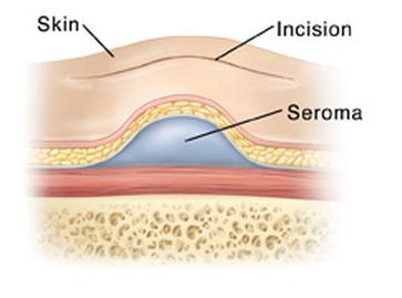
Image 1 – Anatomy of seroma
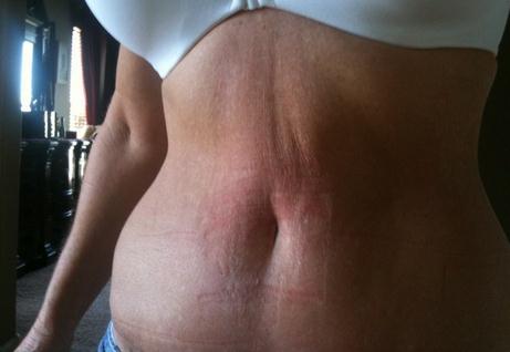
Image 2 – Seroma after plastic surgery (after Tummy tuck)