Archive for November, 2013

Polymyalgia Rheumatica – Symptoms, Diet, Treatment, Causes, Diagnosis

Nov 28 2013 Published by under Diseases & Conditions

What is Polymyalgia Rheumatica?

Polymyalgia Rheumatica, which has an acronym of PMR, is a condition categorized as inflammatory. It is a form of a rheumatic disease linked to stiffness in the shoulder, neck and hip area as well as muscular pain. According to statistics, Polymyalgia Rheumatica affects mostly the elderly and white people. Most often, those who are 65 years old and above are the ones who are inflicted with Polymyalgia Rheumatica. In addition to that, women also reportedly suffer from Polymyalgia Rheumatica more than men.

Polymyalgia Rheumatica Symptoms

Those who suffer from Polymyalgia Rheumatica manifest and report symptoms of painful and severe stiffness upon waking up in the morning and also at night. Most often, the pain and stiffness occurs in the thigh and shoulder. In addition to that, Polymyalgia Rheumatica strikes without prior notice and also appear after illness suhc as the flu.

There are various symptoms that are linked to Polymyalgia Rheumatica. These symptoms include:

  • Mild, low-grade fever
  • Anemia
  • Fatigue
  • Poor appetite
  • Stiffness
  • Headache
  • Blurring of vision
  • Pain in the muscles
  • Depression
  • Weight loss
  • Limited range of motion
  • Edematous distal extremity
  • Having difficulty sleeping
  • Temporal arteritis
  • Synovitis or joint inflammation

polymyalgia rheumatica picture

Neck Pain as a Common Symptom of Polymyalgia Rheumatica

According to studies conducted, persons who suffer from temporal arteritis also have a condition called Polymyalgia Rheumatica and vice versa. Aside from that, those who suffer from Polymyalgia Rheumatica also experience inflammation in their joint area. The classical sign of the Polymyalgia Rheumatica is both stiffness and pain in the muscular region or area.

Polymyalgia Rheumatica Causes

The cause of Polymyalgia Rheumatica is unknown. There has been no evidence of the presence of any toxin or agent that is infectious in nature. The classical signs of Polymyalgia Rheumatica are stiffness and pain which is brought about by the inflammatory process of the body. In, persons suffering from Polymyalgia Rheumatica, the inflammation process is concentrated to the joint tissue area.

According to research, there are a lot of causes or etiological factors, such as:

  • Factors that involves the environment

According to research, the contributing factor may be brought about by the presence of an infectious disease. The presence of a contagious viral diseases may be consistent with new cases of Polymyalgia Rheumatica.

  • Factors that involve genetics

Studies have shown that genes may play a great factor in Polymyalgia Rheumatica. Having a pattern of this disease in the family medical history proves to be evidence that suggests that there is indeed an inheritance influence.

  • Persons with HLA-DR4

HLA-DR4 is a kind of human leukocyte antigen which has a high risk of the getting Polymyalgia Rheumatica.

  • Persons with Giant Cell Arteritis

Polymyalgia Rheumatica also is present in persons affected with Giant Cell Arteritis and vice versa. This kind of condition leads to arterial inflammation.

Polymyalgia Rheumatica Diagnosis

Polymyalgia Rheumatica has no specific and confirmative diagnostic examination. The diagnostic examination given to persons who are suspected of having Polymyalgia Rheumatica will be given a diagnostic examination based on the factors that the person manifests. The diagnostic tests that the person undergoes are the following:

  • Physical examination
  • Medical historical examination
  • Blood examination such as complete blood test which will check for the presence of inflammation
  • Biopsy examination of the temple arteries which will check for the presence of giant cell arteritis
  • Plasma viscosity examination
  • C-reactive protein or CRP examination
  • Erythrocyte sedimentation rate or ESR examination
  • X-ray examination of hips and shoulders
  • Ultrasound examination of hips and shoulders

Polymyalgia Rheumatica Treatment

Persons who suffer from Polymyalgia Rheumatica are given the following suggested treatment options:

Physical therapy

This is an important treatment therapy that is done for the person to regain his or her strength as well as coordination. There is a need for the person with Polymyalgia Rheumatica to be able to undergo physical therapy to be able to perform his or her day to day tasks after being stagnant for a long period..

Medical therapy

Medical therapy is prescribed to persons suffering from Polymyalgia Rheumatica. The medications that are usually prescribed are as follows:

Corticosteroids

Corticosteroid, which is otherwise called glucocorticoid, is a drug classification and the best choice in treating persons suffering from Polymyalgia Rheumatica. The example of medication that belongs to this kind of drug classification is prednisone which is the ideal drug for persons who are inflicted with Polymyalgia Rheumatica. A low dosage of prednisone is given during the initial treatment phase.

Analgesics

The analgesics given to persons with Polymyalgia Rheumatica belong to the classification named NSAIDs or non-steroidal anti-inflammatory drugs like ibuprofen. It relieves the stiffness as well as the pain associated with Polymyalgia Rheumatica.

Bisphosphonates

An example of Bisphosphonates includes alendronate and risedronate which will aid in the prevention of the person to experience osteoporosis.

Polymyalgia Rheumatica Prognosis

The prognosis or outlook for persons that suffer from Polymyalgia Rheumatica is good. Polymyalgia Rheumatica will  resolve after a few years time with the right treatment. Other persons suffering from Polymyalgia Rheumatica may also be relieved of this condition even without undergoing treatment. The treatment mentioned above will only aid in the control of the progression of the disease condition.

Polymyalgia Rheumatica Complications

Persons who suffer from Polymyalgia Rheumatica are at risk of the following complications:

  • Depression
  • Gastrointestinal bleeding due to intake of corticosteroid drugs
  • Temporal arteritis
  • Poor self esteem
  • Physical inactivity

Hence, one must take note of the complication of Polymyalgia Rheumatica.
No responses yet

Wheat Allergy – Symptoms, Diet, Test, Rash, Treatment

Nov 25 2013 Published by under Diseases & Conditions

What is Wheat Allergy?

Wheat allergy is a type of food allergy as a result of ingestion or contact with the different components of wheat. Wheat allergies involve the response of the mast cells and IgE which produces allergy symptoms. Allergies to wheat are commonly due to exposure to the seed storage proteins found in wheat; however, patients may also be allergic to various seeds and plants. There are a total of 27 allergens associated with wheat allergy.

Wheat allergy may be confused with celiac disease and gluten intolerance. The former involves an allergic reaction to proteins in wheat while the latter two cause problems in the intestines as a result of exposure to gluten, a specific protein in wheat.

Symptoms and Signs of Wheat Allergy

Symptoms of wheat allergy are general to all people; however, there are certain symptoms specific to women, adults and children. General symptoms include:

  • Hives

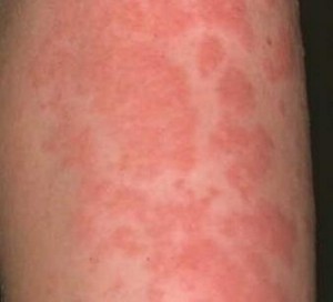
Hives, or urticaria, are raised, bordered, reddish skin patches that are often itchy. They may occur singly or in multiples as a wide reddish patch on the skin. Urticaria is a sign of anaphylaxis and may indicate a life-threatening reaction.

Hives images

Hives

  • Angioedema

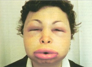
Angioedema is the presence of swelling of the tissues as a result of increased permeability of blood vessels leading to movement of blood from the intravascular space into the interstitial space. Angioedema is mostly observed in the face and leads to swelling of the lips and other tissues.

Angioedema picture

Angioedema

  • Allergic rhinitis

Allergic rhinitis, or hay fever, is the presence of a runny nose along with itchiness and irritation of the nasal passage.

  • Nausea and vomiting

This symptom is a reaction of the body attempting to remove the wheat from the digestive system or inside the body.

  • Abdominal cramps

Allergies to wheat also causes the intestinal mucosa to become irritated, resulting in smooth muscle spasms and causing abdominal cramping.

  • Diarrhea

Diarrhea may also be experienced due to the mechanism of the body to eliminate all foreign substances.

  • Tiredness and lethargy

Allergic reactions to wheat also increase the body’s rate of functioning in order to fight the foreign substance, thereby leading to lack of energy.

Symptoms in women

  • Mood swings

Women may experience changes in mood due to an affectation of the nerve impulse transmission.

  • Hypogastric pain or dysmenorrhea

The presence of swelling of the sacroilial joint may be present as dysmenorrhea when the allergic reaction occurs during menses.

Symptoms in adults

  • Sacroilitis

Sacroilits is the inflammation of the sacroilial joint. Wheat allergies cause the area to swell and inflame as a result of inflammatory response.

  • Eczema

Eczema or atopic dermatitis is the occurrence of rashes on the skin as a result of histamine release by the mast cells, causing vasodilatation and development of scaly rash on the skin. Eczema is often itchy and causes severe dryness of the skin. Eczema can occur in all parts of the body such as the face, arms, back and trunk.

  • Palpitations and chest pain

Adults may experience heart irregularities, especially when there is an anaphylactic reaction or severe allergy. This results from increased heart contractility, resulting in conscious feelings of heart contractions.

  • Joint and muscle pains

Arthritis may also be experienced because of circulation of antibodies to the joints which may lead to damage.

Symptoms in children

  • Unexplained  cough and asthma

Children often exhibit asthma attacks and an unexplained cough. Asthma is more apparent when the child is really asthmatic. Baker’s asthma is a specific allergic reaction to uncooked wheat flour.

  • Swelling of the tongue and throat

There is also pharyngitis that is easily seen in children who suffer from allergic reactions.

  • Wheat allergy rash

Children often do not develop severe eczema. A maculopapular rash may be more apparent in children.

Causes of Wheat Allergy

The main cause of wheat allergy is exposure to different proteins in wheat as a result of ingestion, inhalation or skin contact with these substances. Generally, the main cause is ingestion through food. Foods that contain wheat proteins include:

  • Breads
  • Pastries
  • Cereals
  • Couscous
  • Pasta
  • Spelt
  • Farina
  • Semolina
  • Beer
  • Crackers
  • Soy sauce
  • Vegetable protein
  • Ketchup
  • Hot dogs
  • Ice cream
  • Starch
  • Vegetable gum
  • Jelly beans and candies
  • Licorice

People who are allergic to wheat may also have similar reactions to barley, rye and oats.

Wheat Allergy Testing

Diagnosis of wheat allergy begins with a medical history that might indicate family history of allergies as well as symptomatology. A food diary may also be required to determine the foods that have been taken in. Further testing includes:

  • Skin test

A solution containing small amounts of wheat proteins are injected under the skin to determine if there is an allergy to wheat. The small amount of wheat helps to identify allergy even from the smallest proportion.

  • Antibody testing

Testing for IgE antibodies may also be done to determine presence of immunologic markers for wheat allergy.

  • Food challenge testing

This test involves the ingestion of capsules containing possible allergens. The presence of symptoms following ingestion within hours to days may indicate allergy.

Treatment of Wheat Allergy

The single, most effective way to manage wheat allergy is through avoiding exposure to wheat proteins such as eliminating them from the diet. However, some patients may still ingest portions of wheat in products. With this, specific treatments include:

Medications

Medications such as anti-histamines reduce the symptoms of allergy by binding the histamine receptors to prevent symptoms. Anti-histamines include diphenhydramine, cetirizine, and others. These medications may cause sedation so safety should always be ensured. In cases of anaphylaxis, epinephrine is given to provide emergency management for severe reactions. Epinephrine is usually injected intramuscularly. Patients with allergies are advised to carry epinephrine injections all the time for emergency purposes.

Diet and Recipes

Aside from medications, avoiding the allergen means dietary modifications and restrictions. Wheat products should be avoided. However, lack of wheat intake may also yield deficiencies in fiber. Because of this, wheat-free oats can be used to supply the body with the needed dietary fiber. Recipes that contain wheat-free goodies are also available online. They are easy to prepare and as tasty as foods containing wheat.

No responses yet

Ectodermal Dysplasia (Hypohidrotic) – Pictures, Symptoms, Treatment

Nov 20 2013 Published by under Diseases & Conditions

What is Ectodermal Dysplasia?

Ectodermal Dysplasia is a group of disorders that is congenital and inherited. It affects the tissues of the ectoderm, the outermost layer of the Embryo. A person with Ectodermal Dysplasia has deficiency in the following structures: hair, nails, teeth, and sweat glands. The Ectoderm is involved in developing the nervous system, teeth, epidermis, mucosa, sweat glands, hair and nails.

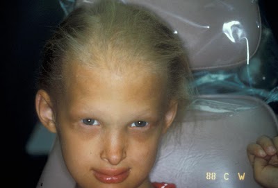
 Hide Ectodermal dysplasia pictures

Ectodermal dysplasia showing fine hair

The first clinical incidence with the same features of Ectodermal dysplasia was during 1792; two Jewish boys had no hair and teeth at birth. At present, there are approximately seven cases per 10,000 affected with Ectodermal dysplasia worldwide. In the United States, there are estimated  cases per 10,000. HED is more common among Caucasians, but there cases come from other races as well.

Types

a. Hypohydrotic

Hypohidrotic ectodermal dysplasia (also known as Anhidriotic ectodermal dysplasia) is the most common type of Ectodermal dysplasia characterized by deficiency in the following:

  • Hair
  • Teeth
  • Sweat glands
  • Mucosa.

There are three inherited types of HED:

  • X-linked recessive HED – the most common type of HED

and the two uncommon type are:

  •  Autosomal dominant HED
  • Autosomal recessive HED.

The incidence of HED is approximately 1 in 10,000 to 1 in 100,000 male live births.

Symptoms

  • General symptoms include:
  • Height that is below expected
  • Repeated acute infectious diseases
  • Fever with unknown reason that occur mostly among infants
  • Hoarseness of voice

Clinical symptoms:

  • Hypodontia (missing teeth)

There is a problem in the development of the teeth resulting in an incomplete number of teeth. Teeth that appear are smaller than the normal size and with cone shaped crowns.

  • Anhidrosis or lack of sweating

Due to malfunctioning of the sweat glands, the body cannot properly control the temperature, thus leading to Hyperthermia or an increase in temperature

  • Hypotrichosis or abnormal hair growth

Growth of body hair is scattered, thin, light in pigment, slow growing, and fragile.

  • Skin

Infections, cracking and bleeding of the skin are common. Unusual pigmentation of the skin is also observed. It can be light pigment, but in some incidence, red or brown pigment occurs.

  • Nails

Abnormalities in the shape of the nails and it affects both the fingernails and toenails. Different color of the nail, slow growing and brittleness are also present.

  • Broader nose

Hollow nasal bridge

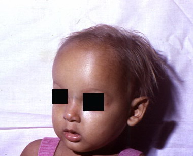
  • Frontal bossing

Abnormal, noticeable forehead.

Frontal bossing Ectodermal dysplasia picture

Frontal bossing in Ectodermal Dysplasia

  • Vision defects

Usually leads to eye infection, cataracts and other eye disorders.

Causes

1. Genetics

Patient siblings

  • X-linked recessive: if the mother is the carrier, there is a 50% chance to be inherited.
  • Autosomal dominant: there is a 50% risk for the siblings to be a carrier; unless the parent is affected, there is no increase of possibilities.

Patients’ offspring

  • X linked recessive

Male proband: 50% of their daughters will be affected and might demostrate minimal symptoms. None of the sons will be the carier.

Female proband: both son and daughter have 50% risk of being a carrier.

  • Autosomal dominant: each child has a 50% risk.
  • Autosomal recessive: there is a 50% risk in the offspring.

Diagnosis

Diagnostic procedures can be done to identify other disorders associated with Ectodermal Dysplasia.

Radiography

Radiographic procedures like Xrays of the extremities can show defect in the bones. Visualization of the kidney through Ultrasound or Intravenous pyelography can assess if the patient is developing anomalies in the urinary system.

Hystopathology

Skin biopsies can identify if there are decrease or absence of sweat and eccrine glands.

Genetic mutation analysis

It is a Prenatal Diagnosis for pregnants carrier of Ectodermal Dysplasia.

Analysis of the Chronic Villi

Treatment

There is no specific treatment to cure Ectodermal dysplasia, but there are many ways to diminish the symptoms.

Temperature

  • Provide a cooling environment inside and outside home with the use of air conditioning, fans and proper ventilation.
  • Drink enough water, especially during the hot season.
  • Wear clothes made of cotton.

Dental management

  • Visit your dentist regularly to monitor the conditions your teeth
  • Wear dentures
  • Dental implantation

Others

  • For dry skin, use moisturizers to prevent eczema and further complications.
  • For the eyes, artificial tears through eye drops can be used to prevent corneal damage.
  • For nasal congestion, you can remove secretions with the use of suctions and humidification of air.
  • Avoid strenuous activities for patients who lack sweat glands (Hypohidrosis).
  • Monitor weight and height.
  • To prevent recurring infections, Antibiotics are given for prophylaxis.
  • Cleft lip or palate repair is done to improve facial appearance and speech.
  • Rehabilation therapy can assist patients in the anomalies they have. Physcial therapy can improve hand and foot abnormalities, speech therapy in speaking and feeding.

Complications

Possible complications include:

  • Febrile seizure due to high fever.
  • Recurrent infections in the upper respiratory tract are common.
  • Inflammation of the scalp that will result to erosion because of Alopecia.
  • Failure in growth development.

No responses yet

Post Inflammatory Hyperpigmentation – Pictures, Treatment, Causes

Nov 14 2013 Published by under Diseases & Conditions

What is Post Inflammatory Hyperpigmentation?

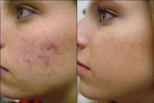
Post Inflammatory Hyperpigmentation is a medical condition that means that the person is suffering from skin discoloration after the recuperation of a skin disease. Other experts would describe Post Inflammatory Hyperpigmentation as a medical term given when the person undergoes skin darkening due to the increased level of melanin which usually is a normal part of the recuperation process. Still others define Post Inflammatory Hyperpigmentation as an acquired illness with the person reporting a presence of skin patches and dark macules at the site of cutaneous inflammatory disease that the person previously experienced. When one has this kind of medical diagnosis, it is widely known medically as the common etiological factors of the alteration of skin color.

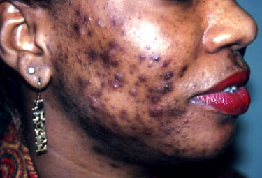
Post Inflammatory Hyperpigmentation picture

Post Inflammatory Hyperpigmentation after acne vulgaris

Post Inflammatory Hyperpigmentation, in addition to that, is given the acronym PIH. Post Inflammatory Hyperpigmentation or PIH is commonly reported to be manifested by persons who have darker skin tones such as those persons belonging to the Mediterraneans, Africa or Asia. Those belonging to these groups are at higher risk of acquiring Post Inflammatory Hyperpigmentation or PIH.

Post Inflammatory Hyperpigmentation Causes

Medically speaking, the etiological causes of why a person undergoes Post Inflammatory Hyperpigmentation are brought about by two medically termed mechanisms which are:

1. Epidermal Melanosis

Epidermal melanosis happens or exists as a result of the release and the oxidation of the arachidonic acid converted to leukotrienes, prostaglandins and the like. This kind of oxidation process is done in a subsequent manner. These are otherwise known as an end product of the inflammatory process which will lead to the alteration of both the melanocyte activity and immune cell activity. Aside from that, they are known to stimulate the epidermal melanocyte which leads to the increase in the melanin synthesis and the increase in the transfer of the pigment that surrounds the keratinocytes. When there is an increase in the stimulation and transfer process of the melanin granules, it will lead to what is medically termed as epidermal hypermelanosis.

2. Dermal Melanosis

Meanwhile, dermal melanosis happens when there is a disruption of the inflammatory process in the person’s basal cell layer which leads to the pigment of the melanin being released and trapped by the macrophages in the person’s papillary dermis which is medically termed as a pigmentary kind of incontinence. Other experts would describe this stage as a deeper pigmentation wherein there is a higher chance that the pigment may be trapped by the tissue macrophages which is a large immune cell.

Post Inflammatory Hyperpigmentation Treatment

With regards to the treatment, it is difficult and a bit prolonged which often takes at a maximum of a year to get the desired results of depigmentation. The treatment management that we mention here will be effective in the improvement of the epidermal hypermelanosis type of Post Inflammatory Hyperpigmentation. However, those who are afflicted with dermal hypermelanosis may not have see an effect with some of the treatments mentioned here. The following are the suggested treatments for persons experiencing Post Inflammatory Hyperpigmentation:

1. Pharmalogical Treatment

The common medical or pharmacological treatments commonly prescribed are topical agents. The topical pharmacological treatments include:

    • Glycolic acid

This is a peel kind of medication which is often called ‘GA peels’ and is used in combination with hydroquinone and Tretinoin. This is an effective pharmacological medication for persons who are suffering from Post Inflammatory Hyperpigmentation and those who have dark skin complexion.

    • Azelaic acid

This is a pharmacological treatment that is used in treating acne vulgaris..

    • Corticosteroids

This classification of medication will target the person’s immune system so as not to target the cascade of the inflammatory process.

    • Tretinoin cream

This pharmacological medication is effective in persons of African American descent. This is a known medical treatment for persons who are suffering from Post Inflammatory Hyperpigmentation.

    • Hydroquinone

This is a commonly prescribed drug for persons suffering from Post Inflammatory Hyperpigmentation. This kind of pharmacological medication is known to be effective in the elimination of the symptoms associated with Post Inflammatory Hyperpigmentation.

Other depigmenting pharmacological agents:

    • Aloe vera leaf extract
    • Glabridin microsponge loaded gel
    • Retinaldehyde
    • Trichloroacetic acid

2. Surgical Treatment

Pertaining to surgical treatment options for persons suffering from Post Inflammatory Hyperpigmentation, the following surgical procedures may be done depending on the assessment done by a physician:

    • Intense pulsed light therapy
    • Chemical peels
    • Laser treatment
    • Fractional photothermolysis
    • 1064-nm Q switched Nd: YAG laser treatment

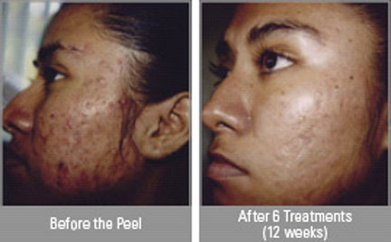
Under the surgical care, we will discuss shortly the two surgical treatment modalities which are laser therapy and chemical peels. A chemical peel can be used as an adjunct in the treatment of Post Inflammatory Hyperpigmentation and those persons who are not responsive to the topical medications prescribed by their respective physicians. On the other hand, laser therapy, which is quite expensive, is not used routinely as it can lead to the person acquiring Post Inflammatory Hyperpigmentation itself. Hence, the risk should be weighed.

Post Inflammatory Hyperpigmentation After Laser treatment pics

Post Inflammatory Hyperpigmentation After Laser treatment

Post Inflammatory Hyperpigmentation picture

Post Inflammatory Hyperpigmentation After Chemical Peels

3. Other treatment options include:

    • Make up that is hypo allergenic in nature

This is a good option that one can use to reduce the effects associated with Post Inflammatory Hyperpigmentation. Aside from that, it will help restore self-esteem during the recuperation stage or process. Also, it is considered to be non-intrusive and a safe treatment option.

    • Natural products

Another treatment option is the use of natural products like herbal extracts and fruit juices which work effectively. An example of natural products includes lemons, papaya, kiwis, grapefruits, raspberry, green tea, mulberry, and licorice which all works to lighten the darkened skin.

No responses yet

White Coating on Tongue – Pictures, Causes, Treatment

Nov 08 2013 Published by under Diseases & Conditions

What is a Coated Tongue?

Coated tongue is a medical symptom that is caused by a bacteria that colonizes in the mouth, leading to halitosis or bad breath which cannot be alleviated, even with mouthwash. Normally, the hue of a person’s tongue ranges from a medium pink to a light pink hue. When a person experiences a coated tongue, the tongue can be either white or yellow. A coated tongue is actually harmless, and it is a symptom, as mentioned earlier, of an underlying illness.

1. White Coated Tongue

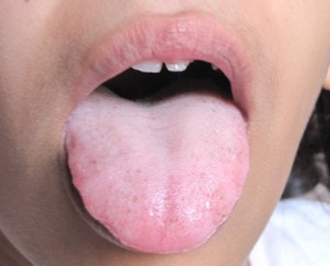
White Coated Tongue Pictures

When a person reports having a white coated tongue, there is no need to panic and cause a scene. The white coated tongue is only temporary and considered harmless. It is often interchanged with the medical term called oral thrush which is caused by an excessive fungal growth in the mouth. When a person usually experiences a white-coated tongue, there are dead cells, bacteria, and debris in the tongue. Moreover, experts say that a white-coated tongue may be due to an underlying health condition which can be infectious or precancerous. In the case of a white-coated tongue, the tongue’s papillae become inflamed due to fever, dehydration, smoking, breathing through the mouth, or other related issues. The bacteria, dead cells, and food debris then become lodged in between the swollen area of the papillae and cause the white-coated tongue symptom.

2. Yellow Coated Tongue

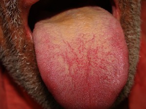
Coated Tongue pictures

A yellow-coated tongue is another problem similar to a white-coated tongue. The main difference is that the former produces a different hue. A yellow-coated tongue, which also occurs from the presence of oral bacteria, is not that grave to cause alarm. Like the white-coated tongue, the yellow-coated tongue happens when the taste buds become swollen, creating a high risk of bacteria build-up. The bacteria that gets lodged between the papillae or taste buds cause the secretion of a yellow-hued chemical which eventually leads to a yellow-coated tongue episode.

What does a Coated Tongue Mean?

Medically speaking, a coated tongue is due to the presence of a white-hued layer composed of bacteria, food particles, and epithelial debris on the uppermost surface of the tongue. Others would define it as a temporary loss of natural tongue color.

What Causes a Coated Tongue?

There are actually a lot of reasons behind coated tongue. Depending on either the white or yellow coated tongue, they have different and some similar etiological reasons behind it. As mentioned earlier, coated tongue may either be white or yellow. The following etiological reasons can predispose a person to experience a white-coated tongue:

  • Smoking
  • Dry mouth
  • Poor oral hygiene
  • Dehydration
  • Bacterial infection
  • herpetic stomatitis or oral herpes virus infection
  • Fever
  • Asthma
  • Diabetes
  • Excessive alcohol consumption
  • Medication side effects
  • Oral thrush
  • Oral lichen planus
  • Syphilis
  • Leukoplakia
  • Candida albicans
  • Map-like appearance or geographic tongue episode

People who suffer from a yellow-coated tongue may show the following symptoms:

  • Heat in the body (part of the inflammatory process)
  • Jaundice (not all yellow coated tongue cases is due to jaundice)
  • Breathing through the mouth
  • Nutritional deficiencies

Coated Tongue Treatments

Normally a yellow or white-coated tongue can lead to social insecurities as well as low self-esteem. The treatments for a white and yellow coated tongue are as follows:

1. Oral health hygiene

This is the best and ideal way to treat white coated tongue to remove bacteria, debris, and the like.

2. Probiotics

This can also be a good way to treat white coated tongue. By squeezing the content from a probiotics capsule onto a spoon and then adding drop of water, the mixture can be swallowed at bedtime.

3. Water consumption

Drinking water provides a lot of heath benefits. For white-coated tongue patients, it will help remove and loosen trapped food debris, bacteria, and dead cells.

4. Smoking cessation

Smoking is one of the causes of white-coated tongues and can lead to other health problems.

5.Healthy and balanced diet

There are many health benefits of a healthy and balanced diet. It is always wise to eat a healthy and balanced diet to prevent nutritional deficiencies and lead a healthy life.

No responses yet

Allergic Contact Dermatitis – Pictures, Symptoms, Treatment

Nov 02 2013 Published by under Diseases & Conditions

What is Allergic Contact Dermatitis?

Allergic Contact Dermatitis is a medical diagnosis in which a person experiences itchy skin that is brought about by an allergic reaction from a certain material coming in contact with the person’s skin. Other medically inclined experts would define it as a contact dermatitis form that has an allergic manifestation response and is due to the irritant substance contact.

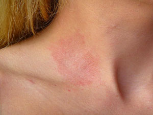
Allergic Contact Dermatitis pictures

Allergic Contact Dermatitis on the side of the neck after wearing jewelry

Medically speaking, this is a prevalent kind of immunotoxicity associated in human beings. Due to the fact that it is allergic in nature, this is a widely known as a hypersensitivity reaction. Hence, it is medically termed as Type IV delayed hypersensitivity reaction which involves the allergic response that is cellular mediated. Aside from that, Allergic Contact Dermatitis is known to rise from the two vital stages known to be the induction phase, where the sensitization is experienced and the other is termed as the anelicitation phase, where the response has been triggered. Most often, the substances that one is allergic to are considered to be harmless such as cotton and the like. Yet, there are persons who have developed hypersensitive reactions to harmless substances.

Allergic Contact Dermatitis Symptoms

Persons suffering from Allergic Contact Dermatitis will experience the following symptoms similar to that of persons suffering from irritant contact dermatitis which leads to a hard diagnosis to begin with. The symptoms from Allergic Contact Dermatitis are as follows:

  • Skin lesions
  • Blisters
  • Vesicles
  • Papules
  • Simple reddish area
  • Itching
  • Inflammation
  • Swelling that is localized
  • Tender to touch
  • Warmth area
  • Leathery skinned
  • Darkened skinned
  • Burning sensation

The first sign that the person with Allergic Contact Dermatitis will observe or notice is a visible red rash. It may not appear for a period of one to two days after being exposed to the irritant. Also, one must know that when the reaction will start; it may take for about a month to completely resolve and return back to normal.

Allergic Contact Dermatitis Causes

Different persons who acquire Allergic Contact Dermatitis will experience different kinds of causes or etiological reasons as to why. The following common causes linked to Allergic Contact Dermatitis are as follows:

  • Exposure to poison sumac
  • Exposure to dyes used in sneakers and shoes
  • Exposure to poison oak
  • Exposure to poison ivy
  • Brought about by newly bought pair of shoes

Generally, exposure to allergens will precipitate the person to experience Allergic Contact Dermatitis. What happens to people suffering from Allergic Contact Dermatitis is that the person’s immune system will react, especially when there is a presence of a foreign substance. The activation of the immune system will lead to the production of the antibodies against allergen that is presence upon contact. This will form a hypersensitivity reaction. The antibody that fights off these foreign invaders are the immunoglobulin E, or IgE, which is stored in mast cells. They are the ones that promote hormonal and chemical release (mediators) such as Histamine. Persons inflicted with Allergic Contact Dermatitis have an external trigger instead of an internal trigger.

Allergic Contact Dermatitis Treatment

With regards to the treatment given to the persons suffering from Allergic Contact Dermatitis:

Pharmacological treatment

The following commonly prescribed treatment medications are as follows:

1. Antihistamine drugs

This may either be prescribed to the person with Allergic Contact Dermatitis or may be bought over-the-counter. This will target the release or activation of the mediator histamine so as not to cause an inflammatory response once there is a presence of allergens or foreign invaders. An example of antihistamine drugs is:

Diphenhydramine medications

This is a known medical treatment that is good for persons experiencing inflammatory reactions. It is commonly prescribed to persons suffering from Allergic Contact Dermatitis.

2. Corticosteroid medications

This drug may either be in a form of an ointment or cream. This works similar to that of the hydrocortisone which is often prescribed to fight off inflammation in a localized area. If there is a severe form of allergy brought about by Allergic Contact Dermatitis, an oral pill or injection may be necessary to assure the direct effect of the medication to the person’s body or immune system.

3. Calamine lotion

This kind of medication may relieve the itchy feeling and may soothe the area that is afflicted by Allergic Contact Dermatitis.

Home remedies

Home remedies include:

  • Draping the affected area in a cool oatmeal bath to aid in the relief of itching episodes
  • Application of wet dressing to the affected area
  • Application of lotion that will dry the skin to avoid itching episodes
  • Wearing smooth textured clothing to prevent irritation of the skin
  • Avoidance of dye
  • Avoidance of perfumes
  • Avoidance of soap that is irritating to the person’s skin
  • Washing the area with cool water after exposure to the allergen
  • Application of cold moist compress especially when blisters are present and applied for as long as 30 minutes for three times a day

Normally, the symptoms associated in Allergic Contact Dermatitis may resolve even without treatment within 2- 4 weeks or one month. As long as the triggering factor or allergen is avoided by the person allergic to it, the recuperation stage will be achieved. However, the person with Allergic Contact Dermatitis may experience a chronic form of the disease condition especially if they were not able to pinpoint the allergen that caused him or her to experience the disease condition. Thus, since it was not pinpointed, it was not avoided.

Allergic Contact Dermatitis Pictures

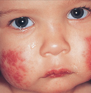
Allergic Contact Dermatitis on the face pics

Allergic contact dermatitis on the face of a child

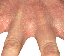
Allergic Contact Dermatitis on the hands pics

Rashes developed after contact with detergent

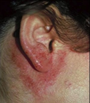
Pictures of Contact dermatitis on the ears

Dermatitis on the ear as a result of frequent contact with hair

No responses yet

© 2017 HealthFoxx.com. All Rights Reserved. Privacy Policy
This website is for informational purposes only and Is not a substitute for medical advice, diagnosis or treatment.