Archive for January, 2014

Idiopathic Pulmonary Fibrosis – Life Expectancy, Treatment, Symptoms

Jan 31 2014 Published by under Diseases & Conditions

What is Idiopathic Pulmonary Fibrosis?

Idiopathic Pulmonary Fibrosis is a delibitating and fatal disease. It is a condition where the lung tissue becomes thickened, stiff, and scarred. The stiffening makes it difficult to breathe because it decreases the elasticity of the lungs due to scar formation. It is an age-related disease that usually affects people between 50-70 years old and the cause is unknown. It is more common in males than females and most of the patients that have Idiopathic Pulmonary Fibrosis have a history of smoking. This condition results from an inflammatory response with an unknown cause. It is a progressive lung disease characterized by formation of fibrosis in the lungs. Fibrosis is a formation of excess connective tissue in a reparative or reactive process: a scar tissue formation in the lungs. As the lungs became scarred, they lose their elasticity and ability to transfer oxygen to the blood stream.

Idiopathic Pulmonary Fibrosis Symptoms

  1. Chest pain
  2. Dry hacking cough
  3. Cyanosis is a condition where it causes bluish discoloration on the skin and mucous membrane.
  4. Decreased tolerance for physical activity due to difficulty in breathing
  5. Shortness of breath during activities or any strenuous exercise
  6. Clubbing of fingers due to lack of oxygenated blood
  7. Crackling sound in the lungs heard through a stethoscope. Crackles are intermittent, discontinuous, explosive, popping sounds. It is heard more during inhalation than exhalation
  8. Fatigue and body malaise because of infection and difficulty breathing
  9. Weight loss
  10. Joint and muscles aches

Idiopathic Pulmonary Fibrosis Causes

The cause for this kind of disease is still unknown. It attacks the lungs, and they will become stiffened, scarred and inflamed. There are some epidemiological factors to be considered such as exposure to (1) cigarette smoking. The damage can be also caused by different things like (2) occupational and environmental factors where there is a long term-exposure to certain toxins and pollutants like asbestos, silica and organic dust. (3) Radiation treatment for cancer can cause lung damage due to exposure to radiation. (4) Medications like chemotherapeutic drugs, heart medications and some antibiotics can cause lung damage. (5) Medical conditions like tuberculosis and pneumonia can affect and cause lung damage; (6) viral or bacterial infection; (7) GERD or Gastro-esophageal Reflux disease where there is a pressure gradient between the abdomen and thorax known to increase negative intra-thoracic pressure associated with the disease that reduces lung compliance. It is a disease of mucosal damage caused by stomach acid coming up into the esophagus.

Diagnosis

a. Medical history and Physical Examination

  1. The doctor will ask you things that can be related into having a Idiopathic Pulmonary Fibrosis such as:
  2.  previous sickness as well as the medications you’re taking.
  3. Physical examination by listening to breathing. Idiopathic Pulmonary Fibrosis is known for abnormal breathing called crackles
  4. If you have a history of smoking.
  5. Your family medical history and how long you have had symptoms.

b. Radiology Diagnosis

  1. Chest X-rays can show the shadow of scar tissues. It isthe  most commonly used diagnostic exam to take images of the lungs and other body parts. It’s a non-invasive examination that helps medical professionals diagnose and treat conditions.
  2. Computed Tomography (CT scan) is a type of X-ray that can provide sharper and more detailed images of the lungs. It combines a series of X-ray views with different angles that produce cross-sectional images of bones and tissues inside the body.

c. Pulmonary Function Test

  1. Spirometry is a breathing test to find out how big is the damage is. It’s a procedure where doctors measure the amount and speed of air during exhalation and inhalation. It can help medical professionals to determine lung disease as well as if medications are working and if the conditions are worsen.
  2. Pulse Oximetry is a gadget that attaches to your ear or fingertips to measure oxygen in the bloodstream. It is not an invasive procedure, just a sensor that attaches to the body to measure arterial blood.
  3. Exercise Stress Test using treadmill or stationary bike monitors lung function. It is a useful way of evaluating symptoms of shortness of breath, stridor or wheezing during exercise.

d. Tissue Sample

  1. Biopsy is a medical test performed by a surgeon to get a sample tissue for examination. It can determine the presence and extent of the disease. It can help the doctors diagnose the exact problem in the patient.
  2. Video Assisted Thoracoscopy is also a procedure to get tissue samples using an endoscopic tube with a camera and light attached to it.

Idiopathic Pulmonary Fibrosis Treatment

There is still no known cure for Idiopathic Pulmonary Fibrosis. The goal of treating Idiopathic Pulmonary Fibrosis is to reduce further scarring which is irreversible, relieve symptoms like difficulty of breathing, and improve or maintain quality life. The treatments only focus in alleviating pain and disturbances that affect a persons daily routine.

  1. Medications such as corticosteroids and cytotoxic drugs help to reduce inflammation as well as N-acetylcysteine, an anti-oxidant that prevents lung damage
  2. Oxygen therapy can’t stop lung damage but it can help alleviate difficulty of breathing and prevent or lessen complications of low-blood oxygen levels. It helps to reduce breathlessness and helps the patient become more active.
  3. Lung Rehabilitation is a standard care given to patients that have a lung problem. It allows the person to manage their condition and improve their quality of life. It may include breathing exercises, nutritional counseling, support groups, education about the disease, how to manage it, energy-conserving techniques, breathing strategies and anxiety, stress, or depression management. It will help the patient to function better. Support groups are there so patients can ease the stress of having this disease and where patients can share common experience and problems.
  4. A lung transplant is recommended if the condition worsen. It can improve quality of life as well as help you live longer.

Prognosis

It depends on the progression of the disease. Some cases may stop progressing; some may develop slowly or quickly into the end-stage disease. Some patients may end up with supplementary oxygen and some require lung transplants. The course of this disease is difficult to predict; however, the average survival time for an individual with Idiopathic Pulmonary Fibrosis is 5-7 years.
No responses yet

Epidermal Inclusion Cyst – Causes, Treatment, Diagnosis

Jan 27 2014 Published by under Diseases & Conditions

What is an Epidermal Inclusion Cyst?

Epidermal Inclusion Cysts otherwise known as epidermoid cysts, epidermal cyst, epidermal inclusion cyst, infundibular cyst and keratin cyst is a widely found cutaneous cyst. Smaller than 10% of epidermal inclusion cysts happen in the extremity and an even lesser degree occurs in the palms, fingers, toes, sole, and breast. The record shows that it is commonly found in the skin layer of the face, scalp, neck, and trunk.
The name epidermal inclusion cyst means embedded epidermal elements in the dermis. This is because most of these vesicles attached in the infundibular portion of the hair follicle. In most cases, epidermoid cyst is the preferred term.

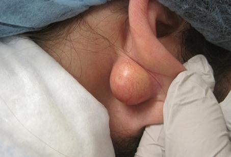
epidermal inclusion cyst

Epidermoid cysts are usually without obvious symptoms. An epidermal cyst is either fixed or movable lump with “cheese-like” material lying under the skin. This cyst is usually benign and the size is approximately 8mm. It is rare to spread, or grow fast and unlikely to bleed. It does not hurt upon palpation and not secured to deeper structures. In the event of inflammation although atypical, results in pain and tenderness.

Causes of epidermal inclusion cyst

Age

Epidermal cysts may exist at any age range; yet commonly appear between 30 to 40 years old. On the other hand, small epidermoid cysts called milia are common in newborn children.

Sex

Epidermoid cysts are nearly double in male than in female.

Trauma

Repetitive irritation, blunt trauma and crushing injuries take part in the transformation of epidermoid cysts. The period between trauma and the development of a cyst may range from a few months to 22 years.

Surgery

Invasive procedures such as rhinoplasty, breast augmentation, reduction mammoplasty, liposuction, dermal grafts, myocutaneous grafts, and needle biopsies subsequently result in epidermoid cysts.

UV

The harmful effects of ultraviolet light may play a role in the development of epidermoid cysts. This condition is referred to as a Favre-Racouchot syndrome.

Others

Congenital

Sequestered epidermal cells during embryonic life may cause epidermoid cysts.

Hereditary

Some heritable disorder is associated with epidermoid cysts particularly an autosomal dominant disease called Gardner syndrome.

Idiopathic

A medical condition called scrotal calcification contains epidermoid cysts which occur without apparent cause.

Race

Race has not been well established in this disorder. Epidermoid cyst is believed to be common in a distinct group of people with dark skin. In fact, a research of the Indian population with epidermoid cysts proved otherwise. It turned out 63% of the cyst had melanin substance.

HPV

Experts consider human papillomavirus (HPV) to cause epidermoid cysts though unclear.

Acne

Acne formation can lead to a follicular occlusion. Documented cases are when the hair follicles or pores are impeded and inflammation and growth occur in the lower level of the epidermis made an inclusion cyst.

Diagnosis

A medical history and physical examination will be obtained from a specialist in skin disorders called Dermatologist. The recommendation is to undergo:

Imaging studies

The procedure listed below is advisable when an epidermoid cyst believed to be situated in uncommon position. Such locations are breast, a section of the skeleton, or within the skull.

  1. Ultrasonography
  2. Radiography
  3. CT scans
  4. MRI

Fine-needle aspiration

To diagnose epidermoid cysts in the breast, aspiration is performed. Then the aspirated material is stained with Wright-Giemsa solution. Positive results will show nucleated keratinocytes and curl keratin material.

Microscopic studies

Epidermoid cysts are consisting of stratified squamous epithelium with a granular layer. Histology study will also show keratin contents inside the cyst. On the other hand, calcification may be displayed in older cysts.

Laboratory Studies

In cases of repeated infection or absence of significant response to antibiotic therapy, a culture and sensitivity may be obtained.

Another test

Biopsy is not necessary if common sonographic and physical examination findings are found. Nonetheless, in cases showing palpable breast lesions, the patients are often concerned about lumps and may ask for excision.

Treatment of epidermal cyst

Epidermoid cysts with no symptoms do not need to be treated. However, treatment may include the following:

Medical intervention

Those suffering from inflammatory response may benefit from antibiotics administered by mouth. Alternatively, corticosteroid reduces swelling. Cysts displaying no inflammation and no infection may benefit to an intralesional injection of triamcinolone.

Surgical Care

Through the wonders of surgery, getting rid of epidermoid cysts would be fast and easy. It is done by simple excision or incision of the cyst and cyst wall. It is very important to remove the whole cyst or else it will recur. Extraction of cyst surgically with the punch biopsy procedure may be used in ideal locations. Moreover, decreased possibility of scarring has been documented in small-incision surgery.

Surgical drainage may be carried out if a cyst is infected. This is to speed up healing; although, it will not remove the cyst.

Complications of epidermal cyst

Preventing an epidermal cyst is nearly impossible but complications are infrequent. These include:

  1. Infection
  2. Spontaneous rupture of the cyst

These cysts emit nonabsorbable keratin base fluid that acts as an irritant causing a secondary foreign body-type response and granulomatous reaction.

  1. Formation of abscess
  2. Chances of long incision and high possibility of scarring
  3. Recurrence

If any of the cyst walls is left behind after incision and drainage, the cyst may develop again.

Carcinoma

Malignancies in epidermoid cysts are very unusual. But there is a record showing basal cell carcinoma, Bowen disease, squamous cell cancer (SCC), metaplastic lesions including mycosis fungoides, a rise in epidermoid cysts.

Paget’s disease

Some experts have documented cases of Paget’s disease arising from the epidermal inclusion cyst.

No responses yet

Heel Pain – Causes, Treatment, Relief, Types, Diagnosis

Jan 26 2014 Published by under Diseases & Conditions

What is Heel Pain?

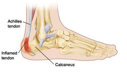
Heel pain is a condition in which weight-bearing on the heel causes discomfort. It i a common foot problem where pain usually occurs underneath or behind the heel where the Achilles tendon attaches to the bone.

Heel Pain pictures

Types of Heel Pain

a. Achilles Tendinitis

  1. An inflammation of the tendon that connects calf muscles to the heel bones.
  2. Overuse of Achilles tendon can cause inflammation, pain, and swelling.
  3. It is caused by overuse of the affected limb and this condition is most common in people who engage in exercises like jogging, running or who play tennis.
  4. The Achilles tendon is considered the largest tendon in the body which primarily used for running, walking and jumping.
  5. The Achilles tendon doesn’t have good blood supply, so when injury occurs, it heals slowly.
  6. Cause: It is more likely to occur after an increase in intensity or amount of activity such as playing basketball and (b) when the calf muscles are not stretched.
  7. Symptoms: (a) pain in the back of the heel which usually worsens with activity (b) pain and stiffness along the Achilles tendon in the morning (c) severe pain the day after exercising (d) thickening of the tendon as a result of swelling (e) painful when touched or moved (g) swollen and warm to touch (f) tenderness, especially during palpation
  8. Diagnosis: (a)  history of prior injuries (b) physical examination: (1)the doctor will palpate the location of the injury and look for signs of tenderness, swelling, and pain in the area of tendon when you stand in your toes. (2) The doctor will also check for passive and active range of motions and reflexes of the knees, ankle and feet. (c) Imaging test like X-rays can show if the tendon have become calcified or hardened, and magnetic resonance imaging (MRI) can help look for tears within the tendon.
  9. Treatment: (a) cold compress (b) wearing heel pads to reduce stress in the tendon (c) an exercise routine to help strengthen the tendon (d) rest to prevent the situation becoming worse
  10. (e) NSAIDS or non-steroidal anti-inflammatory drugs like ibuprofen to reduce inflammation and pain (f) manual therapy that treats musculoskeletal pain or injury comprised of massage therapy, and joint mobilization and manipulation. (g) If these treatments do not improve symptoms, surgery is recommended to remove bone spur that irritate the tendon. (h) Extracorporeal shock wave therapy that uses low dose of sound waves to stimulate the healing process in the damaged area. It is not an invasive procedure, so it is recommended before surgery, but it doesn’t have a consistent result.

b. Bursitis of the heel

  1. It is also called Retrocalcaneal bursitis
  2. Is an inflammation of the bursa, a small sack of fluid beneath the heel bone.
  3. A bursal sac is filled with slippery fluids that acts to lubricate and helps reduce friction between the muscles and the tendons. It acts as a cushion or shock absorber between tendons and the bone.
  4. It is located between the Achilles tendon and the heel bone.
  5. Cause: (a) when a person increases their level of physical activity like too much walking and running, it irritates and inflames the bursa (b) over-using ankles by doing repetitive actions (c) ill-fitting shoes (d) or medical problems like rheumatoid and gouty arthritis.
  6. Symptoms: (a) pain in the heel especially when running or walking, and even when the area is touched (b) extreme pain especially when standing on tiptoes (c) tender feel when it is palpated (d) warm to the touch and redness the skin over the back of the heel.
  7. Diagnosis: (a) prior injury history (b) physical examination that involves checking the infected part for pain, redness, swelling, and warmth (c) imaging tests like X-rays which can show if the tendon has become calcified or hardened, and magnetic resonance imaging (MRI) which can help evaluate a patient for tears within the tendon.
  8. Treatment: (a) manual therapy treats musculoskeletal pain or injury and is comprise of massage therapy, joint mobilization and manipulation (b) activity modification: avoid activities that cause pain (c) cold compress (d) exercise that will improve strength, flexibility and balance (e) NSAIDS or non-steroidal anti-inflammatory drugs like ibuprofen to reduce inflammation and pain (f) ultrasound treatments help reduce the inflammation (g) normally this type of disease doesn’t require surgery

c. Excessive Pronation

  1. Pronation is a normal adaption of the foot to adapt on uneven surfaces while walking and it also helps absorb shock.
  2. Excessive pronation is where the foot turns inward and soft tissue stretches and causes stress and inflammation on the planter fascia ligament.
  3. Cause: (a) very common for flat-footed persons (b) trauma from sports or repeated stress from wearing high-heeled shoes for long periods of time (c) shoes lacking adequate support through the arch
  4. Symptoms: (a) Achilles tendinitis (b) bunion (c) heel spurs (d) plantar fasciitis (e) ankle sprain (f) knee, hip, lower back pain (g) corns and calluses (h) hammertoes or contracted toes which is a result of wearing ill-fitting shoes; the toes became bent like a hammer (i) shin splints or lower leg pain
  5. Diagnosis: (a) history of prior injury (b) physical examination checking the infected part for pain, redness, swelling, and warmth of the heel area (c) there is a test to detect excessive pronation: (1) the Achilles tendon test where the person will stand in front of the mirror and look at the back of the legs and foot. On a normal foot, the tendon and the heel run straight down, but in excessive pronation, the tendon runs straight down the leg but twist outwards at the heel. (2) A “used shoes” test where you look at your used running shoes with the toes pointed away from you; if your shoes tilt inward, it shows that your feet are excessively pronated.
  6. Treatment: (a) anti-pronation insoles provide a supprt system that helps aligns the lower body (b) wearing supportive shoes that give ample support and cushioning particularly at the heel and arch of the foot (c) surgery is not that reccomended in this type of heel problem

d. Haglund’s Deformity

  1. Known medically as posterior calcaneal exotosis that is a bony enlargement surrounded by tender tissue on the back of the heel of the bone.
  2. The soft tissue near the Achilles tendon becomes irritated, especially when it rubs against the shoes.
  3. It commonly called “pump bump” because of the high-heeled shoes that women usually wear.
  4. Cause: (a) high-heeled shoes or any shoes with a rigid back can cause irritation (b) heredity also plays a role in this disease (1) a high-arched foot means the heel of the bone is tilted backward into Achilles tendon and this causes the upper portion of the heel to rub against the tendon (2) a tight Achilles tendon can cause pain by compressing the bursa and a tendency to walk outside the heel that produces wear on the outer edge of the sole of the shoes that causes the heel to rotate inward.
  5. Symptoms: (a) a noticeable bump on the back of the heel (b) pain in the area where the Achilles tendon is attached (c) swelling, redness and tenderness on the back of the heel
  6. Diagnosis: (a) history of prior injury (b) physical examination checking for pain, redness, swelling, and warmth of the heel area (c) imaging tests like X-rays which evaluate the structure of the heel bone, size of the deformity and the involvement of the Achilles tendon
  7. Treatment: (a) ice compression (b) change of footwear (c) use of Achilles heel pads that may help reduce irritation when walking (d) NSAIDS or non-steroidal anti-inflammatory drugs like ibuprofen to reduce inflammation and pain (e) stretching exercises that will help the Achilles tendon reduce tension (f) immobilization if necessary (g) surgery is considered in severe cases to remove or reduce the bony growth

e. Plantar Fasciitis and Heel Spur Syndrome

  1. The plantar fascia works like a rubberband between the heel and the ball of the foot to form and support the foot’s arch. This is also a shock absorber for the body.
  2. It is a painful condition often caused by overuse of the plantar fascia.
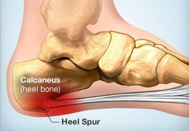
  3. Heel spurs are a pointed bony fragment that stems from the bone and when the plantar fascia is overused, it will form calcium deposits under the heel bone as a result of inflammation. Heel spurs don’t causes pain but they are indicators that the person may suffer from Plantar fasciitis.
  4. Cause: (a) a common cause is very tight calf muscles that result in over-pronating that causes additional stretching of the plantar fascia (b) heredity is also included by having a high or low-arched foot (c) you walk or run on hard surfaces without enough support (d) being overweight will put strain on your heel (e) shoes that don’t fit well or are worn out (f) long-distance running
  5. Symptoms: (a) pain at the bottom of the foot near the heel (b) pain after exercising or any strenuous activity (c) pain is worse when you take the first step in the morning or after a long period of rest (d) sudden stretching of the foot may make the pain worse (e) after standing or sitting
  6. Diagnosis: (a) history of prior injury (b) physical examination checking for pain, redness, swelling, and warmth of the heel area (c) imaging tests like X-rays or an ultrasound that shows thickening or swelling of the fascia
  7. Treatment: (a) rest (b) massage therapy (c) stretching (d) weight loss (e) motion control shoes or shoes with thick insoles and shoes that have extra cushioning (f) warm and cold therapy (g) NSAIDS or non-steroidal anti-inflammatory drugs like ibuprofen to reduce inflammation and pain (h) stretching exercises to relieve pain (i) using extracorporeal shock wave therapy that uses low dose of sound waves to stimulate the healing process; it is not an invasive procedure so it is recommended before surgery, but it doesn’t have a consistent result (j) surgery is considered if the pain is not eased for 12 months despite other treatments

No responses yet

Baker’s Cyst – Pictures, Symptoms, Treatment, Surgery

Jan 20 2014 Published by under Diseases & Conditions

What is Baker’s Cyst?

Baker’s cyst, also known as popliteal cyst or bulge-knee, is a swelling of the synovial bursa located behind the knees (popliteal area). The swelling comes from synovial fluid in the knee joint that accumulates at the concave area behind the knee. The term ‘baker’s cyst’ was named for a British doctor named William Morrant Baker, the surgeon who first described it. It is a benign condition and not actually a “cyst” because it is not encapsulated and open communication with the synovial sac is preserved.

Baker’s cyst in adults usually develop due to excess weight that compresses the synovial fluid, leading to protrusion at the back of the knee. When the bulge grows, it becomes palpable.

Baker’s Cyst picture

Ruptured Baker’s Cyst

A baker’s cyst, when constantly irritated, can rupture. Constant irritation may include heavy lifting, running, climbing, squatting and kneeling despite the presence of baker’s cyst. The continued pressure on the knee joint and bursa causes the baker’s cyst to rupture. This causes protrusion of fluid down the leg between the gastrocnemius muscles (calf muscle) and leads to dissection. The leak of fluid down the inner leg causes symptoms such as:

  1. Appearance of bruising on the calf and inner ankle.
  2. Rapid swelling of the leg and calf muscle
  3. Acute pain behind the knee and calf muscle
  4. Redness in the calf

The symptoms of a ruptured baker’s cyst are similar to the symptoms of thrombophlebitis and the more serious deep vein thrombosis (DVT). These conditions cause thrombus formation on the veins that may be dislodged, causing life-threatening embolism, stroke, and myocardial infraction. For these reasons, ruptured baker’s cysts should be determined from thrombophlebitis and deep vein thrombosis for immediate management of said conditions. This is done through diagnostic tests such as ultrasounds and blood tests. Immediate medical treatment is required for deep vein thrombosis.

Baker’s cyst may also compress blood vessels and cause edema of the legs. Injury to blood vessels causes an inflammatory reaction and clot formation that may potentially lead to deep vein thrombosis. Although this is an infrequent occurrence, baker’s cyst should be managed early to prevent such complications.

The fluid from the ruptured cyst is often systematically absorbed by the body, but this does not mean that the baker’s cyst will go away. Usually, the swelling comes back and may lead to complication of the baker’s cyst again.

Ruptured baker’s cysts are usually treated with corticosteroid injections in the knee, rest and leg elevation. Arthropscopic surgery may also be done to permanently remove the baker’s cyst.

Causes of Baker’s Cyst

Baker’s cyst is caused primarily by swelling of the synovial joint that results from increase in synovial fluid due to certain factors. The swelling in turn increases pressure inside the bursa (the sac that contains the synovial fluid), causing the area to bulge at the back of the knee or popliteal area.

The most common reasons for swelling include:

Osteoarthritis

Osteoarthritis or degenerative arthritis causes irritation and compression of the synovial sac, causing it to bulge. This is the most common cause of baker’s cyst in adults.

Juvenile arthritis

Arthritis that occurs at a young age also causes swelling of the synovial bursa.

Cartilage Tears

Tears in the leg such as a torn meniscus (tears on the cartilage of the knee) and rheumatoid arthritis also may lead to baker’s cysts.

Symptoms of Baker’s Cyst

Baker’s cyst in some individuals is asymptomatic although some may also experience symptoms. The most common symptoms associated with baker’s cyst include the following:

  1. Tightness behind the knees when the knees are fully flexed or extended
  2. Knee pain, especially after movement or exercise
  3. Visible bulge behind the knee, especially when standing
  4. Soft and tender bulge, likened to a water-filled balloon
  5. Swellings the size of a golf ball
  6. Pressure that may extend into the calf muscle
  7. Limited range of knee motion
  8. Red glow on the area using transillumination (shine a light on the bulge with lights turned off)

Ruptured baker’s cyst can also cause significant pain, swelling, and bruising on the calf and inner ankle.

Diagnosis of Baker’s Cyst

Diagnosing baker’s cyst begins with physical examination of the knees, popliteal area, and calf muscles. Palpation is done to characterize the bulge and determine the presence of tenderness and pain.

Diagnostic tests are also employed, including:

Ultrasonography

Ultrasound of the popliteal area is the most common test for baker’s cyst to determine the extent of the swelling.

Magnetic Resonance Imaging

MRI can also reveal the presence of swelling in the popliteal area as well as any meniscal injury or tear.

Arthrogram

This involves the injection of dye into the knee, followed by imaging test.

Transillumination

Shining light on the bulge can show the presence of fluid, manifested by a red glow.

X-ray

X-rays are used to determine the presence of other problems such as arthritis.

When the bulge occurs with bizarre signs such as a rapid increase in size, sever pain during daytime and nighttime, fever, and difficulty of movement, the presence of tumors should be ruled out by doing more diagnostic tests.

Treatment of Baker’s Cyst

Asymptomatic baker’s cysts usually do not require any treatment. Baker’s cysts in children also do not require invasive managements. When symptoms occur, it’s better to manage the case to reduce uncomfortable feelings. Treatment for baker’s cyst involves medical and surgical treatment, natural remedies, and exercise.

Medical Treatment

  1. Corticoseroids. Corticosteroids are injected into the area to reduce swelling and inflammation
  2. Analgesics and anti-inflammatory medications. These are given to reduce inflammation and pain. Be sure to take the medication after meals to reduce gastrointestinal irritation.

Natural Treatment

  1. Ice packs. Ice packs are placed over the area to reduce swelling and numb the area to reduce pain. Ice packs should be applied ten to fifteen minutes to achieve better results. This should be done during the acute phase of the condition.
  2. Heat. Warm compresses over the area are helpful during chronic pain and bruising. This reduces pain by relaxing the structures on the area and also decreases the blood clot as a result of ruptured cysts.
  3. Rest. Avoid strenuous activities that involve the knees and legs to prevent further injury. These activities may include running, squatting, climbing, kneeling, and lifting heavy objects.

Physiotherapy

  1. Strengthening of the muscles. Exercises that focus on strengthening the calf and knee ligaments may help reduce symptoms and improve mobility. Stretching and other isometric and isotonic exercises are often suggested by a physiotherapist.
  2. Bracing. Knee braces are used to support the joint and prevent further injury. In mild cases, a support using elastic bandage can be used.

Aspiration

Bakers’s cysts are usually resolved by aspirating the excess fluids in the area. Costicosteroids are usually injected after the procedure.

Surgery

Knee surgeries are often done with cases of other internal knee problems such as cartilage tears. The procedure involves arthroscopic surgery where the surgeon removes the swollen synovium. By removing this, the cyst formation is prevented from reoccurring. Arthroscopic surgery uses a special scope to visualize the internal aspects of the knee joint that aids the surgeon in doing the surgery.

Recovery from the procedure usually takes eight to twelve weeks. During this time, excessive motion of the knees should be prevented, although a simple range of motion exercises is essential in restoring the mobility of the knee joint.

Complications of Baker’s Cyst

Complications of baker’s cyst do not usually develop if treatments are implemented. Uncommon complications include:

  1. Meniscal tears
  2. Rupture of the baker’s cyst
  3. Thrombophlebitis
  4. Bruising
  5. Chronic pain and swelling

Prognosis

Baker’s cysts do not cause significant harm if measures are taken. However, it can be painful and irritating. The symptoms of baker’s cyst are also easy to manage and most people recover after arthroscopic surgery. Long-term disability and complications are usually rare.

Since it is a benign condition, it usually does not spread or cause malignancy.

No responses yet

Pre Menopause – Symptoms, Signs, Diet, Treatment

Jan 14 2014 Published by under Diseases & Conditions

Pre menopause is the time or stage in life prior to menopause. It is known as perimenopause, or the phase when women experience changes in the reproductive functioning before a complete cessation of menses. There is also cessation of egg production by the ovaries during menopause. Because of this, a woman may no longer menstruate. The term ‘pre menopause’ may cause misconceptions as pertaining to children or adolescence and the period before menopause. Because of this, gynecologists and most physicians accept the term perimenopause.

Pre menopause Age

The average age for pre menopause starts beteen 35 to 40 years of age. During these times, women experience signs of changing hormone levels. However, there is no specific time when pre-menopause may occur because each woman has unique characteristics and may experience symptoms differently.

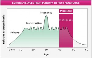
Pre-Menopause Hormones pic

The Estrogen Levels throughout Womanhood with the levels decreasing during Menopause

Symptoms and signs of Pre Menopause

There are a lot of symptoms that may be experienced during the pre menopause stage. All of these are associated with the reduction of hormone levels in women. Symptoms and signs include:

  • Irregular periods

When the hormones are already reduced because of pre menopause, there will be irregularities in menstruation due to the absence of adequate hormones to regulate the menstrual cycle. Irregular periods may occur from four to ten years prior to complete cessation of menses. Irregular menses may mean shorter or longer periods than before as well as lighter or heavier menses than normal. The perimenopause stage is considered over when the woman experiences menopause by not having menses for one year or more.

  • Irritability and Mood Swings

Estrogen and progesterone also regulate the mood of a person. The scenario is similar to the premenstrual syndrome women experience. Prior to menstruation, the level of the hormones decrease which leads to mood swings. Women may need emotional and psychological support during this stage in order for them to cope with the changes in their bodies. Lack of support may lead to depression

  • Decrease in Bone Density

Estrogen hormones reduces bone resorption or the escape of calcium in the bones. When estrogen levels decrease, regulation is limited, thereby allowing the reduction of calcium in bones and in blood, leading to a reduction in the bone density. This explains why older women are more prone to osteoporosis.

  • Hot Flashes

Hot flashes are also a common symptom. The condition appears as a sudden increase in body temperature. Hot flashes can cause discomfort, especially when there is a sudden increase in body temperature of more than two degrees. Hot flashes are the inability of the body to regulate temperature efficiently. During hot flashes, women may experience intense sweating and feelings of weakness or fatigue similar to fever. However, hot flashes are not considered harmful and may resolve in a few minutes or hours.

  • Weight Gain

Women may also experience weight gain because of water retention. This happens due to the inability of the body to regulate water.

  • Breast Tenderness

Breast tenderness is also experienced similar to the premenstrual syndrome as a result of the reduction in estrogen levels.

  • Thinning of Hair and Drying of Skin

Women may also experience changes in the integumentary system because of erratic changes in hormone levels.

  • Migraine Headaches

Headaches are a common symptom of the reduction in hormone levels.

  • Decreased Libido

Sex hormones are also responsible for the sexual functioning of women. Changes lead to decreased libido and sexual desires.

  • Insomnia

Insomnia may also be experienced as a result of psychological disturbances and hot flashes at night.

  • Loss of Vaginal and Urinary Tone

A decrease in estrogen leads to loss of elasticity and lubrication of the vagina. This may cause increased susceptibility to infections and painful sexual intercourse. This also leads to loss of tone of the urethral meatus and urinary incontinence.

  • Increase in Cholesterol Levels

Once estrogen has decreased, there will be an increase in the bad cholesterol and a reduction in good cholesterol that may predispose women to certain heart diseases.

Treatment Regimen for Pre-menopause

Prior to the onset of menopause, women should consult a gynecologist in order to adapt to menopause easily. Certain therapies are employed to help the woman undergo the transition in her life without severe discomforts. Therapies include:

Hormone replacement Therapy

The basic treatment is to replace the hormones in the body which have decreased a result of pre menopause. Hormone replacement therapy may include oral contraceptives which have estrogen content or a woman may choose progestin therapy. When hormones are established, they help women return to their normal body function and reduce most symptoms.

Endometrial ablation

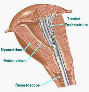
Endometrial ablation may be used for women suffering from heavy menstrual flow. This procedure involves the destruction of the thick endometrial lining with the use of heat, electrical energy or laser.

Endometrial Ablation procedure pics

Endometrial Ablation Procedure Involving Scraping the Thick Endometrail Lining

Proper nutrition

Calcium is very essential for pre-menopausal women to reduce the risk for osteoporosis. A low-fat diet may also be practiced to prevent high cholesterol levels and to prevent heart diseases.

Stress Reduction activities

Pre menopausal women may experience stress as a result of discomfort associated with the transition. Women should engage in activities like yoga, meditation, massage therapy or other stress-relieving therapies to help them cope.

Exercise

Exercise is also essential to maintain healthy bones and overall health. Adequate exercise also prevents weight gain and improves the sleep pattern of pre menopausal women.

Alternative medicine

Women may also consider alternative medicines such as the use of a back cohosh, phytoestrogens and DHEA. Black cohosh is an herb extract that is beneficial for the management of hot flashes and other symptoms. Phytoestrogens are compounds found in plants such as flax seeds that are similar to natural estrogen in the body. Finally, dehydroepiandrosterone (or DHEA) is a steroid produced by the adrenal gland to reduce symptoms of pre menopause. DHEA can be taken in the form of supplements.

No responses yet

Bartholin Gland Cyst – Pictures, Causes, Treatment

Jan 06 2014 Published by under Diseases & Conditions

What are Bartholin Glands?

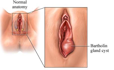
The Bartholin glands are two pea-sized glands located on the either side of the vaginal orifice under the skin. Bartholin glands are small enough which makes it non-palpable. One cannot see the Bartholin glands directly from the vulva because they are located under the tissues. Bartholin glands are also termed as Greater vestibular glands.

Bartholin glands were first described by a Danish anatomist, Caspar Bartholin the Younger during the 17th century. It was the discovery of the young Bartholin and not of his grandfather, Caspar Bartholin the Elder.

Functions

The Bartholin glands are essential during sexual intercourse. These two glands secrete mucus to lubricate the vagina during intercourse. It lubricates the outer genital area with fluids that come out of the Bartholin ducts into the opening of the vagina.

The mucus secreted by the Bartholin glands are relatively small and about one to two drops only. It is secreted when the woman is sexually aroused. The fluid also moistens the labia minora to make contact to the area less uncomfortable for the woman.

Bartholin gland Pathology

The Bartholin glands are sensitive to irritation and infection. Certain factors cause the ducts to become obstructed. When this happens, cysts may form and become infected. Infection of the glands and ducts may lead to abscess formation.

Cancer, tumors and hyperplasia of the Bartholin glands are rare.

What is Bartholin gland Cyst?

Obstruction in the duct causes fluid to build up because it is unable to be released to the vaginal opening. The formation of obstruction and fluid accumulation is termed as Bartholin gland cyst or Bartholin duct cyst.

Cysts that arise on the area are usually the size of a pea or marble. However, it can grow and form an abscess as a result of infection. Bartholin cyst are also usually painless and only one of the two ducts become affected.

Symptoms

Uncomplicated Bartholin gland cysts have the following symptoms:

  1. Painless lump on the vulva near the vaginal orifice
  2. Swelling and redness in the area
  3. Cyst may vary in size from a quarter of an inch to one inch. The cyst usually enlarges during sexual intercourse as a result of more fluid accumulating on the area.
  4. Dyspareunia (painful sexual intercourse). The woman may feel pain during intercourse as a result of irritation of the cyst and the absence of adequate lubrication resulting from the obstructed duct.

If Bartholin’s cysts become infected, the woman usually manifests the following symptoms:

  1. Pain. Severe pain on the cysts may be experienced and may result in difficulty walking and sitting.
  2. Abscess formation. Formation of pus in the area is a sign of an ongoing infectious process.
  3. Tenderness. Tenderness may be experienced on the surrounding areas of the cyst.
  4. Vaginal Discharges. Abnormal discharges may be present if the cyst is caused by a sexually transmitted disease.
  5. Fever. Fever is a sign of infection and some women may experience it.

Causes

Causes of Bartholin’s gland cysts vary, but they usually result from obstruction of the duct. These include:

Infection

Sexually transmitted diseases such as Chlamydia and gonorrhea cause the duct to become infected. Infection results in production of discharges that potentially blocks the duct. As a result, it causes fluid build-up and cyst formation.

Bacteria found on the intestinal tract such as Escherichia coli may also infect the duct and cause cyst formation.

Poor Personal Hygiene

Because of poor hygiene, infection may occur in the ducts and glands. Poor hygiene after sexual intercourse significantly increases chances of Bartholin’s gland cysts.

Use of restrictive pants and undergarments.

Restrictive clothing does not allow circulation of air and blood in the area and leads to blockage and cyst formation.

Diagnosis

Diagnosis of Bartholin’s gland generally involves physical examination of the vulva. Uncomplicated or uninfected cysts do not require special diagnostic tests.

In cases of infection, cultures and sensitivity testing of the abscess are done to determine the specific type of bacteria and the antibiotic treatment to be used. Smears of the vagina and cervix may also be done to determine presence of sexually transmitted diseases.

Treatment

Women with Bartholin gland cysts often seek treatment as a result of pain and difficulty walking or sitting. Some cyst resolve themselves, but others do not. Treatment of Bartholin cyst involves removal (surgery) and antibiotic use. Home remedies and prevention may also be instituted.

Antibiotic Treatment

Bartholin’s gland abscess requires antibiotic treatment to eradicate the causative microorganism. Antibiotics are in the form of penicillinns, cephalosporins or macrolides.

Hot sitz bath

Hot sitz bath involves the exposure of the vulva to hot steam to allow the abscess to be drained by itself. This also reduces pain sensations and hastens healing because of vasodilatation in the area that allows better blood circulation. This can be done at home by sitting in a tub with warm water for 15 minutes three times a day. Be sure not to use hot water because it may burn the skin and cause more damage.

Marsupialization

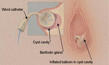
Marsupialzation involves a minor surgery to drain the abscess and remove the cyst. This is often done in an out-patient setting and does not require hospital admission. The procedure is done first by injecting local anesthetics around the surgical site. A small incision is made to drain the fluid and abscess. Next, the cyst wall is sutured to the skin to prevent the cyst from reccurring.

Marsupialization is often uncomfortable because local anesthetics often do not act well on the area. Depending on the choice of the patient and physician, sedation may be used.

Analgesics

Pain medications are usually prescribed to reduce pain as a result of the cyst or after surgical removal.

Prevention

Preventing cyst formation entails the following measures:

  1. Lower the risk for sexually transmitted diseases by using barrier protection such as condoms.
  2. Practice safe sex by practicing monogamy to prevent STDs
  3. Practice good perineal hygiene. These include perineal care, change of underwear, and change of napkins or tampons every two hours.
  4. Avoid using non-restrictive garments to allow for better circulation.
  5. Use cotton under garments. Cotton absorbs secretions well and prevents bacterial growth unlike satins and silk.

Complications

Complications do not usually develop; however, an infected cyst that is not treated may spread to underlying structures and may cause more extensive infection. Unremoved cysts through surgical treatment also often reoccurs. Malignancies are uncommon with Bartholin’s gland cysts.

Pictures

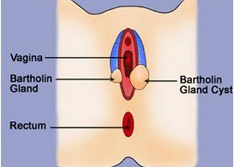
Bartholin gland cyst image

Image 1 – Bartholin Gland Cyst

Bartholin's gland cyst

Image 2 – Bartholin’s Gland cyst image

Bartholin Gland Cyst drainage

Image 3 – Bartholin Gland Drianage

No responses yet

Calcaneal Apophysitis – Symptoms, Causes, Treatment

Jan 01 2014 Published by under Diseases & Conditions

What is Calcaneal Apophysitis?

It is otherwise known as Sever’s Disease which is an inflammation of the person’s calcaneal growth plate or “apophysis” which is located at the posterior part of the calcaneus, the medical term for the heel bone. This leads to disability as well as pain. It affects mostly children, often boys, between the ages of 8-14. Children of this age are highly affected because the heel bone or “calcaneus” is not yet developed fully until the child reaches 14 years of age when there will be recurrent stress on the growth plate and it can lead to inflammation. It can occur in both or one foot.  It is different from heel pain that adults experience which may improve in time because the Sever’s disease doesn’t subside or doesn’t improve.

Calcaneal Apophysitis Image

Calcaneal Apophysitis Symptoms

Persons with Calcaneal Apophysitis will experience the following symptoms associated with the condition, such as:

  • Episodes of limping
  • Walking on toes only
  • Difficulty engaging in an active lifestyle that requires jumping, running, or other sports-related activities
  • Pain, especially when the affected heel is squeezed
  • Pain in the bottom of the affected heel
  • Tenderness in the affected area
  • Swelling in the affected area

Calcaneal Apophysitis Causes

As mentioned earlier, the calcaneal Apophysitis occurs most often in children who overuse their heel bone or have undergone recurrent trauma of the heel bone such as sprain injuries, occurring most often in sports. Aside from that, having a congenital tight muscles in the calf is a high risk for developing this condition. Calcaneal Apophysitis happens because:

  • Trauma such as a sprain or injury
  • Overused heel bone
  • Congenital tight muscles in the calf

Calcaneal Apophysitis Diagnosis

Orthopedic professionals are the ones who diagnose children with this kind of condition. Prior to diagnosing, they require them to undergo certain tests to confirm that they really have this condition. Such tests include:

  • Medical history assessment test
  • Physical assessment test
  • X-ray
  • Magnetic Resonance Imaging or MRI

These are the basic and common tests that a patient undergoes to confirm this condition. However, the physician may require other tests to rule out other possible conditions.

Calcaneal Apophysitis Treatment

When it comes to treating the patient diagnosed with calcaneal Apophysitis, the treatment will vary from one person to another. The treatment will be individualized and may require one of the following:

Heel support

Custom orthotic devices or shoe inserts that may be used temporarily are good ways to provide heel support.

Shoe Modification

Wear shoes that lessens stress on the affected heel bone. Aside from that, it is suggested that the patient wears shoes that have an open back to reduce the pressure of the affected heel bone. Also, wear a higher-heeled shoe. Wearing such will lead to the elevation of the heel, decrease the force required, and allow pain to subside gradually.

Reduction of activity

When a patient, specifically a child, has this kind of condition, he or she is advised to stop all the activities which may lead to pain

Pharmacological treatment

The physician usually prescribes medications of NSAIDs or non-steroidal anti-inflammatory drugs like ibuprofen which acts to reduce the inflammation as well as pain.

Immobilization

When the person is dealing with the severe form of this disease condition, he or she should immobilize the affected heel through the use of a cast. This is done to aid in recuperation of the affected tissue.

Physical therapy

In promoting healing, there is a need for physical or stretching therapy. It promotes healing as it aids in blood circulation.

Ice application

Through ice application for at least 20 minutes maximum, it will reduce inflammation, swelling, and cool the area that is affected.

This kind of condition may recur while the bone is still immature. When this happens, contact a trusted ankle and foot surgeon as soon as possible.

No responses yet

© 2017 HealthFoxx.com. All Rights Reserved. Privacy Policy
This website is for informational purposes only and Is not a substitute for medical advice, diagnosis or treatment.