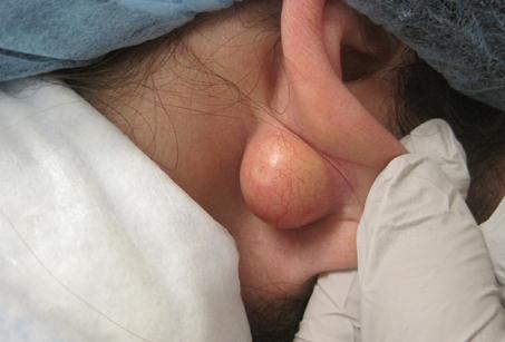
Epidermal Inclusion Cyst – Causes, Treatment, Diagnosis
What is an Epidermal Inclusion Cyst?
Epidermal Inclusion Cysts otherwise known as epidermoid cysts, epidermal cyst, epidermal inclusion cyst, infundibular cyst and keratin cyst is a widely found cutaneous cyst. Smaller than 10% of epidermal inclusion cysts happen in the extremity and an even lesser degree occurs in the palms, fingers, toes, sole, and breast. The record shows that it is commonly found in the skin layer of the face, scalp, neck, and trunk.
The name epidermal inclusion cyst means embedded epidermal elements in the dermis. This is because most of these vesicles attached in the infundibular portion of the hair follicle. In most cases, epidermoid cyst is the preferred term.

Epidermoid cysts are usually without obvious symptoms. An epidermal cyst is either fixed or movable lump with “cheese-like” material lying under the skin. This cyst is usually benign and the size is approximately 8mm. It is rare to spread, or grow fast and unlikely to bleed. It does not hurt upon palpation and not secured to deeper structures. In the event of inflammation although atypical, results in pain and tenderness.
Causes of epidermal inclusion cyst
Age
Epidermal cysts may exist at any age range; yet commonly appear between 30 to 40 years old. On the other hand, small epidermoid cysts called milia are common in newborn children.
Sex
Epidermoid cysts are nearly double in male than in female.
Trauma
Repetitive irritation, blunt trauma and crushing injuries take part in the transformation of epidermoid cysts. The period between trauma and the development of a cyst may range from a few months to 22 years.
Surgery
Invasive procedures such as rhinoplasty, breast augmentation, reduction mammoplasty, liposuction, dermal grafts, myocutaneous grafts, and needle biopsies subsequently result in epidermoid cysts.
UV
The harmful effects of ultraviolet light may play a role in the development of epidermoid cysts. This condition is referred to as a Favre-Racouchot syndrome.
Others
Congenital
Sequestered epidermal cells during embryonic life may cause epidermoid cysts.
Hereditary
Some heritable disorder is associated with epidermoid cysts particularly an autosomal dominant disease called Gardner syndrome.
Idiopathic
A medical condition called scrotal calcification contains epidermoid cysts which occur without apparent cause.
Race
Race has not been well established in this disorder. Epidermoid cyst is believed to be common in a distinct group of people with dark skin. In fact, a research of the Indian population with epidermoid cysts proved otherwise. It turned out 63% of the cyst had melanin substance.
HPV
Experts consider human papillomavirus (HPV) to cause epidermoid cysts though unclear.
Acne
Acne formation can lead to a follicular occlusion. Documented cases are when the hair follicles or pores are impeded and inflammation and growth occur in the lower level of the epidermis made an inclusion cyst.
Diagnosis
A medical history and physical examination will be obtained from a specialist in skin disorders called Dermatologist. The recommendation is to undergo:
Imaging studies
The procedure listed below is advisable when an epidermoid cyst believed to be situated in uncommon position. Such locations are breast, a section of the skeleton, or within the skull.
- Ultrasonography
- Radiography
- CT scans
- MRI
Fine-needle aspiration
To diagnose epidermoid cysts in the breast, aspiration is performed. Then the aspirated material is stained with Wright-Giemsa solution. Positive results will show nucleated keratinocytes and curl keratin material.
Microscopic studies
Epidermoid cysts are consisting of stratified squamous epithelium with a granular layer. Histology study will also show keratin contents inside the cyst. On the other hand, calcification may be displayed in older cysts.
Laboratory Studies
In cases of repeated infection or absence of significant response to antibiotic therapy, a culture and sensitivity may be obtained.
Another test
Biopsy is not necessary if common sonographic and physical examination findings are found. Nonetheless, in cases showing palpable breast lesions, the patients are often concerned about lumps and may ask for excision.
Treatment of epidermal cyst
Epidermoid cysts with no symptoms do not need to be treated. However, treatment may include the following:
Medical intervention
Those suffering from inflammatory response may benefit from antibiotics administered by mouth. Alternatively, corticosteroid reduces swelling. Cysts displaying no inflammation and no infection may benefit to an intralesional injection of triamcinolone.
Surgical Care
Through the wonders of surgery, getting rid of epidermoid cysts would be fast and easy. It is done by simple excision or incision of the cyst and cyst wall. It is very important to remove the whole cyst or else it will recur. Extraction of cyst surgically with the punch biopsy procedure may be used in ideal locations. Moreover, decreased possibility of scarring has been documented in small-incision surgery.
Surgical drainage may be carried out if a cyst is infected. This is to speed up healing; although, it will not remove the cyst.
Complications of epidermal cyst
Preventing an epidermal cyst is nearly impossible but complications are infrequent. These include:
- Infection
- Spontaneous rupture of the cyst
These cysts emit nonabsorbable keratin base fluid that acts as an irritant causing a secondary foreign body-type response and granulomatous reaction.
- Formation of abscess
- Chances of long incision and high possibility of scarring
- Recurrence
If any of the cyst walls is left behind after incision and drainage, the cyst may develop again.
Carcinoma
Malignancies in epidermoid cysts are very unusual. But there is a record showing basal cell carcinoma, Bowen disease, squamous cell cancer (SCC), metaplastic lesions including mycosis fungoides, a rise in epidermoid cysts.
Paget’s disease
Some experts have documented cases of Paget’s disease arising from the epidermal inclusion cyst.